
Skip to newsletter content

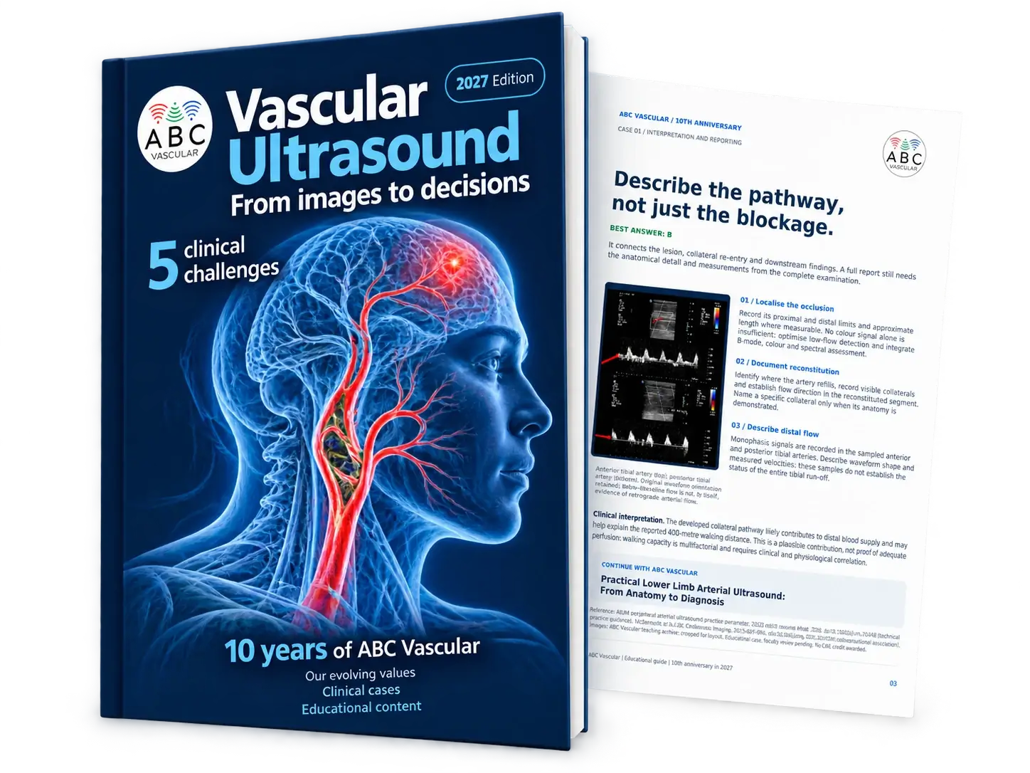
Free eBook 2027 edition

From images.
To decisions.

Five clinical challenges.
One free guide to vascular ultrasound.

Clinical case studiesFrom images to interpretation
Illustrated explanationsImages, interpretation, reporting
ABC Vascular expertiseEducation for clinical practice
Subscribe & get eBook Take a look inside

Clinical cases.
Practical questions.
Clear explanations.

Vascular Ultrasound, From images to decisions: 2027 eBook with an open sample page.
2027 editionComplimentary PDF

Inside the guide

What would you report?

Explore real pages from the guide. Five challenges connect ultrasound
findings with interpretation and reporting.

Pages 3–4 of the guide · Select a page to enlarge

Read this caseLower limb arterial

The question and explanation from the selected guide preview, in text.

Page 3

CASE 01 / LOWER LIMB ARTERIAL ULTRASOUND

SFA occlusion:

What should your ultrasound report include?

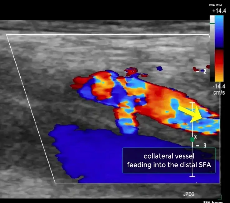
B / Distal reconstitution. A collateral vessel is shown entering the distal SFA.

Which report best communicates the findings?

A. SFA occlusion. Distal arteries patent.

C. Chronic SFA occlusion with adequate collateral perfusion; no further haemodynamic assessment is needed.

Clinical history. The patient reports a walking distance of approximately 400 metres. Duplex assessment identifies SFA occlusion with distal collateral reconstitution.

A / Occluded segment. No colour filling is shown in the sampled SFA segment after lowering the colour velocity scale.

These selected images illustrate the occluded segment and distal collateral re-entry. A complete report should map the lesion and describe downstream haemodynamics.

B. SFA occlusion with collateral reconstitution of the distal SFA and monophasic flow in the sampled tibial arteries; document the occlusion extent, re-entry site and distal assessment.

Page 4

CASE 01 / INTERPRETATION AND REPORTING

Describe the pathway, not just the blockage.

BEST ANSWER: B

It connects the lesion, collateral re-entry and downstream findings. A full report still needs the anatomical detail and measurements from the complete examination.

01 / Localise the occlusion Record its proximal and distal limits and approximate length where measurable. No colour signal alone is insufficient: optimise low-flow detection and integrate B-mode, colour and spectral assessment.

02 / Document reconstitution Identify where the artery refills, record visible collaterals and establish flow direction in the reconstituted segment.

Name a specific collateral only when its anatomy is demonstrated.

03 / Describe distal flow Monophasic signals are recorded in the sampled anterior and posterior tibial arteries. Describe waveform shape and measured velocities; these samples do not establish the status of the entire tibial run-off.

Anterior tibial artery (top); posterior tibial artery (bottom). Original waveform orientation retained.

Below-baseline flow alone is not evidence of retrograde arterial flow.

Clinical interpretation The developed collateral pathway likely contributes to distal blood supply and may help explain the reported 400-metre walking distance. This is a plausible contribution, not proof of adequate perfusion: walking capacity is multifactorial and requires clinical and physiological correlation.

CONTINUE WITH ABC VASCULAR

Practical Lower Limb Arterial Ultrasound:

From Anatomy to Diagnosis Images extracted from ABC Vascular master courses.

Page 5

CHALLENGE 02 / CAROTID PLAQUE CHARACTERISATION

Beyond the stenosis:

How would you characterise these plaques?

Compare plaque morphology and explain the different clinical pathways.

PLAQUE A

ASYMPTOMATIC SCENARIO

A 68-year-old patient has an incidental carotid plaque and no previous carotid-territory symptoms.

In this fictional scenario, ICA stenosis is 40% by diameter reduction using NASCET.

PLAQUE B

SYMPTOMATIC SCENARIO

A 72-year-old patient develops transient left monocular visual loss.

YOUR CHALLENGE

1. How do echogenicity, internal texture and surface differ?

2. Which features suggest a potentially higher-risk phenotype?

3. Why would the two clinical pathways differ?

In this teaching scenario, urgent assessment identifies a left ICA lesion with 80-90% stenosis by NASCET criteria.

Images extracted from ABC Vascular master courses. Clinical histories are constructed teaching scenarios.

Plaque B stenosis (80-90% NASCET) is supplied by the author, not measured from this still image.

Page 6

CHALLENGE 02 / INTERPRETATION AND CLINICAL PATHWAY

Characterise first.

Interpret in context.

PLAQUE A / A less concerning appearance PLAQUE B / Features that raise concern

HOW THE PATHWAYS DIFFER IN THESE SCENARIOS

DEVELOP YOUR PLAQUE ASSESSMENT SKILLS

Carotid Atherosclerotic Plaque Assessment Explore the ABC Vascular Master Course > Images extracted from ABC Vascular master courses.

A / Medical management. The asymptomatic patient with 40% diameter reduction using NASCET receives vascular risk assessment, lipid-lowering treatment, appropriate antithrombotic therapy and risk-factor management. Surgery is not indicated by the image alone.

The learning point. Characterisation can refine risk assessment and strengthen a clinically useful report. Symptoms, validated stenosis grading, additional imaging and patient factors determine management together; plaque morphology is not a stand-alone indication for surgery.

The plaque has a relatively smooth luminal contour in this view. Describe its echogenicity and texture alongside the surface. A smooth appearance alone cannot establish biological stability or exclude future events.

This transverse image shows a predominantly anechoic plaque component. Describe its echogenicity and assess the plaque in multiple optimised views. B-mode alone cannot confirm a lipid-rich necrotic core, intraplaque haemorrhage or cap rupture. The 80-90% NASCET grade is supplied by the author; it cannot be established from this still image alone.

B / Endarterectomy plus medical management. Following urgent stroke and vascular assessment, the patient with recent ipsilateral symptoms and 80-90% stenosis by NASCET criteria undergoes carotid endarterectomy within 14 days, after assessment of surgical suitability and procedural risk.

Medical treatment continues alongside surgery.

Page 7

CHALLENGE 03 / ICA STENOSIS GRADING

One artery.

Five different PSVs.

Which stenosis grade would you report?

SIMULATED ICA MEASUREMENTS / SAME SAMPLE LOCATION

BEAT 1

160

BEAT 2

205

BEAT 3

245

BEAT 4

185

BEAT 5

225 PSV in cm/s. Highest beat: 245 cm/s. Five-beat mean: 204 cm/s.

What is the most defensible response?

A. Report 70-79% NASCET stenosis because one PSV exceeds 230 cm/s.

B. Report 50-69% stenosis because the five-beat mean is 204 cm/s.

Clinical scenario. A 74-year-old patient with known atrial fibrillation undergoes carotid duplex assessment. Plaque narrows the ICA lumen, and the spectral recording shows marked variation between cardiac cycles.

The available recording does not include reliable ICA EDV or a CCA reference acquisition. Plaque and colour images suggest narrowing, but no complete anatomical or haemodynamic assessment is available.

C. Complete a protocol-consistent assessment, integrate the parameters and state the effect of arrhythmia before assigning a grade.

Teaching scenario. The patient and five measurements are simulated. They illustrate the reasoning problem and are not extracted from an ABC patient recording. NASCET refers to diameter reduction, not area reduction.

Page 8

CHALLENGE 03 / INTEGRATED INTERPRETATION

The grade needs the complete assessment.

BEST ANSWER: C

Atrial fibrillation changes acquisition

WHAT THE COMPLETE ASSESSMENT SHOULD CONNECT

LEARN THE FULL SCANNING AND GRADING APPROACH

Carotid and Vertebral Arteries Master Course Explore the course >

SIMULATED DOPPLER ENVELOPE

Irregular intervals; variable peak velocities The highest beat and the arithmetic mean answer different measurement questions. Neither, on its own, establishes the stenosis grade in this incomplete AF assessment.

Record several cycles and document the rhythm.

Where possible, sample later beats in a run of relatively regular cycles. If your protocol uses averaging, state the method. Avoid selecting an isolated unusually high or low beat.

Technique and haemodynamics: verified flow alignment and sample position, representative ICA PSV/EDV and distal CCA PSV/EDV, ratios and spectral findings. Apply additional indices, such as the St Mary's ratio, only within the validated local criteria.

Anatomy and circulation: plaque, residual lumen, distal ICA calibre, contralateral disease and vertebral findings. Suspected near-occlusion requires specific assessment: velocities may be high, low or undetectable. Describe plaque risk features separately from stenosis severity.

Schematic illustration of beat-to-beat variation in AF. Simulated values; not a patient recording.

EDV and ICA/CCA ratios also vary. Use comparable acquisition conditions: a high ICA beat divided by a low CCA beat can create a misleading ratio.

Page 9

CHALLENGE 04 / ABDOMINAL AORTIC ANEURYSM

A giant AAA.

What exactly are you measuring?

Which measurement should your report describe?

A. The diameter of the patent flow lumen only.

Case finding. A giant fusiform infrarenal abdominal aortic aneurysm with mural thrombus and a reported maximum AP diameter of 10.0 cm, measured outer-to-outer.

B. The maximum AP diameter across the whole aneurysm sac, from the outer anterior wall to the outer posterior wall, in an appropriate orthogonal plane.

C. The longest oblique diameter visible on any single image, regardless of the aortic axis.

Image extracted from the ABC Vascular AAA master course. Maximum AP diameter: 10 cm outer-to-outer (case measurement).

Page 10

CHALLENGE 04 / MEASUREMENT AND REPORTING

The sac diameter needs a defined method.

BEST ANSWER: B

OUTER-TO-OUTER AP

Anterior outer wall Posterior outer wall Teaching diagram; not to scale.

Find the maximum AP diameter Place calipers on the outer walls Keep the convention consistent

MEASUREMENT, CLASSIFICATION AND REPORTING

Abdominal Aortic Aneurysm Master Course Explore the ABC Vascular course > Images extracted from ABC Vascular master courses.

Sweep the aneurysm and identify its maximum AP dimension. Align the measurement plane perpendicular to the local longitudinal aortic axis. Oblique sections may exaggerate the diameter.

For this outer-to-outer measurement, include both walls and the whole sac, including any mural thrombus. Measuring only the patent lumen underestimates the aneurysm diameter.

State the plane and caliper method.

Inner-to-inner, leading-edge and outer-to-outer measurements are not interchangeable. Use the same protocol for valid serial comparisons.

Suggested report wording: "Giant fusiform infrarenal AAA with mural thrombus. Maximum AP diameter 100 mm (10.0 cm), measured outer-to-outer in a plane perpendicular to the aortic axis." Use this wording once the full examination confirms these details.

Shape and thrombus also matter. Report fusiform or saccular morphology and the presence and distribution of mural thrombus. Saccular AAAs may rupture at smaller diameters and warrant individual specialist assessment; evidence is mainly observational. Mural thrombus does not establish protection from rupture. This 100 mm AAA requires urgent vascular assessment; new pain or haemodynamic instability requires emergency assessment.

Page 11

CHALLENGE 05 / VARICOSE VEINS

Before you treat, build the map.

Anatomy first. Reflux second. A report that guides planning.

A / Transverse B-mode anatomy of the GSV B / Provoked spectral Doppler at the SFJ Your scanning challenge Before reading the answer, identify what you still need:

1. The course of the GSV, its tributaries and perforator connections.

2. Where reflux starts, how far it extends and which segments remain competent.

3. The anatomy and haemodynamics needed for an individual treatment plan.

A reflux finding is the beginning of the map, not the end of the examination.

Clinical history. A 31-year-old man with primary right-leg varicose veins reports heaviness, ankle swelling and itching. Varicosities extend along the medial calf. No previous DVT or venous surgery is reported.

You detect reflux at the saphenofemoral junction (SFJ). Does this mean the entire great saphenous vein (GSV) should be considered incompetent?

Page 12

CHALLENGE 05 / STEP 1 - B-MODE ANATOMY

First, know which vein you are following.

Build an anatomical route before interpreting flow.

01 / Identify the junction 02 / Follow the GSV 03 / Follow the varicosities 04 / Identify perforators Recognise the SFJ and common femoral vein.

Assess deep-vein patency and compressibility; complete the survey of other relevant trunks.

Trace the medial-calf varicosities to their connection with the GSV. Record course, distribution and tortuosity.

Trace the vein in its fascial compartment.

Distinguish the saphenous trunk from a superficial tributary.

Locate fascial crossings and deep connections.

Record diameter and location; size alone does not establish incompetence.

Page 13

CHALLENGE 05 / STEP 2 - TRACE THE REFLUX

Where does reflux start, and where does it stop?

Test the junction, the trunk and the connected branches.

01 / Provoke and measure 02 / Test the distal GSV 03 / Assess the perforator Assess standing whenever possible. Provoke flow at the SFJ and measure reverse-flow duration. Baseline polarity depends on orientation and inversion settings.

Use distal compression-release or cuff deflation.

Identify the transition to a competent segment; do not extrapolate from a proximal result.

Interpret the complete pattern The teaching case describes SFJ and thigh-GSV incompetence, calf varicosities and a competent distal GSV. A medial-calf perforator is also described as incompetent.

Common thresholds: >0.5 s in superficial truncal veins; >1 s in the CFV, femoral and popliteal veins. Use your validated protocol.

Confirm flow direction and reflux duration with spectral Doppler. Diameter or a colour flash alone cannot establish incompetence.

Page 14

CHALLENGE 05 / STEP 3 - MAP AND REPORT

A map the treating clinician can use.

ANSWER: SFJ REFLUX DOES NOT MAKE THE WHOLE GSV INCOMPETENT

Describe the pattern, not just “reflux”.

Complete the treatment-planning report

LEARN THE FULL VENOUS ASSESSMENT

Lower Limb Venous System Course Explore the ABC Vascular course > Measure diameter for procedural planning Images extracted from ABC Vascular master courses.

Right SFJ and thigh GSV reflux, associated with medial-calf varicosities. The GSV is described as competent distal to their origin. An incompetent medial-calf perforator is reported in the teaching acquisition.

Add patient position, provocation manoeuvres, reflux durations, segment lengths and diameter sites. Include deep-system patency and reflux, tributary connections, perforator location and direction, vein depth, tortuosity and any technical limitations.

Measure the anteroposterior diameter of truncal veins in cross-sectional view. A diameter >3 mm is generally needed in some EVLT protocols; technical suitability depends on the device and access anatomy. This is not a universal treatment threshold.

Diagram from the ABC Vascular master course. Red: incompetent pathway. Blue:

competent distal GSV. Not to scale.

Why it matters: map refluxing targets and competent segments separately. An incompetent perforator is not automatically a treatment target. Decisions integrate the scan, clinical assessment and patient preferences.

Stay connected with ABC Vascular

Keep learning beyond the eBook.

Get practical vascular ultrasound insights delivered to your inbox.
Choose the updates that matter to your clinical practice.

Inside the 2027 guide

Enlarged guide pageSubscribe & get eBook
New Release

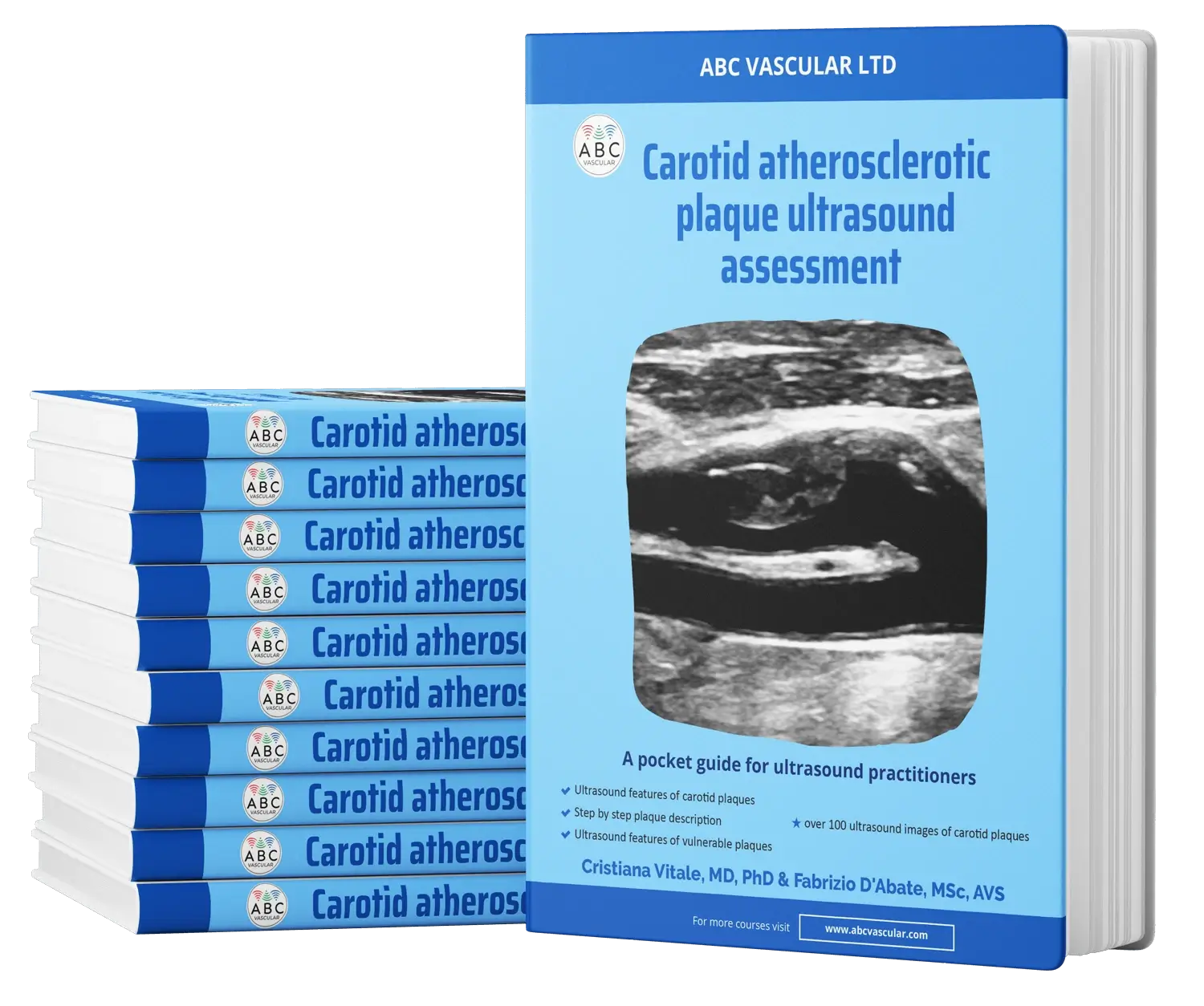
Your Ultimate Guide to Carotid plaque Ultrasound Assessment

By: C. Vitale & F. D'Abate

Explore the world of carotid atherosclerotic plaques with ABC Vascular’s latest eBook, “A Practical Guide on the Ultrasound Assessment of Carotid Atherosclerotic Plaques”. This guide offers healthcare professionals a comprehensive understanding of carotid plaque ultrasound assessment and its role in cardiovascular risk management.

Trusted by