FREE EBOOK · 2027 EDITION
Five Clinical Challenges.
One Free Illustrated Guide.
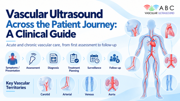
Join the ABC Vascular newsletter and receive our free 2027 eBook, “Vascular Ultrasound: From images to decisions”.
Deep Vein Thrombosis: Ultrasound Criteria & Diagnostic Skills for Healthcare Professionals
A practical guide to the direct and indirect ultrasound findings of lower limb DVT, compression techniques, Doppler interpretation and common diagnostic pitfalls.

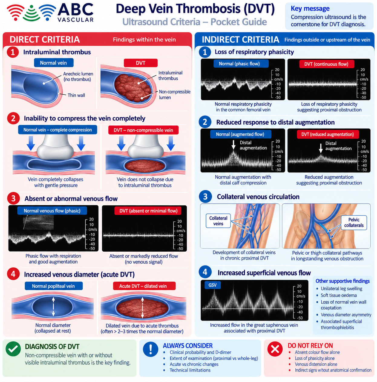
Deep vein thrombosis (DVT) occurs when thrombus forms within the deep venous system, potentially impairing venous return and exposing patients to complications including pulmonary embolism. Ultrasound is the first-line imaging investigation for suspected lower limb DVT, but an accurate examination requires systematic scanning, appropriate technique and interpretation within the clinical context.
In this guide, we review the direct and indirect ultrasound criteria used to assess DVT, explain their relative diagnostic importance and highlight when a negative or technically limited examination requires further consideration.
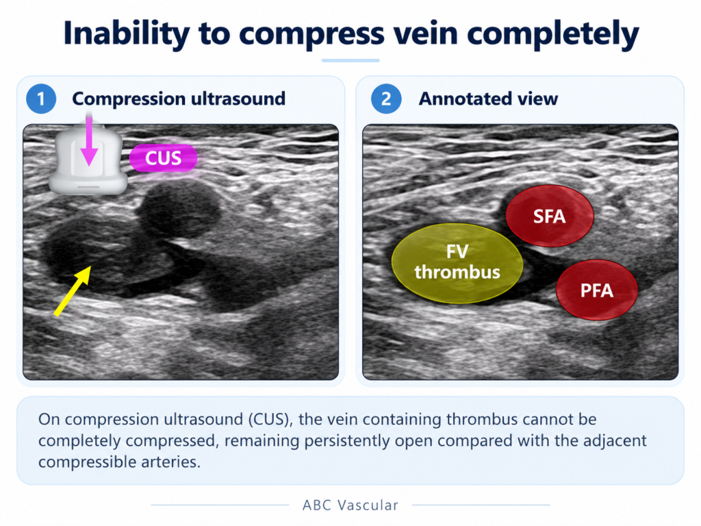
With gentle, correctly directed transducer pressure, a normal vein should collapse until its anterior and posterior walls meet. Failure of complete coaptation is the central direct sign of DVT. Compression must be assessed in an appropriate transverse plane with the entire venous segment adequately visualised.
Apparent non-compressibility can arise from technical factors such as insufficient pressure, excessive depth, adjacent tissue or patient discomfort. Residual post-thrombotic changes can also affect venous compressibility, so ultrasound appearances must be interpreted with previous examinations and clinical history when available.
Figure 1. Compression ultrasound and failure of complete venous collapse. Educational composite; final scientific review should confirm the illustrated anatomy and labels before publication.
Build confidence in recognising the main direct diagnostic criteria through commented scanning demonstrations, structured learning material and a CME quiz.
Explore the DVT Short Course →A focused introduction to DVT diagnosis. Full course details and eligibility are available on the course page.
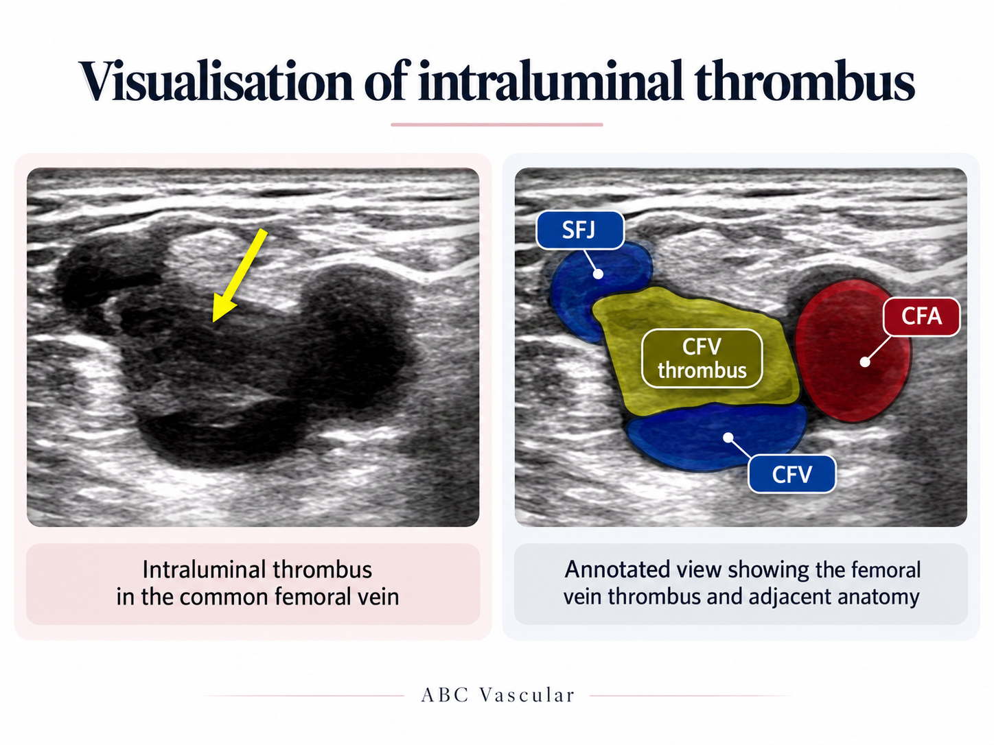
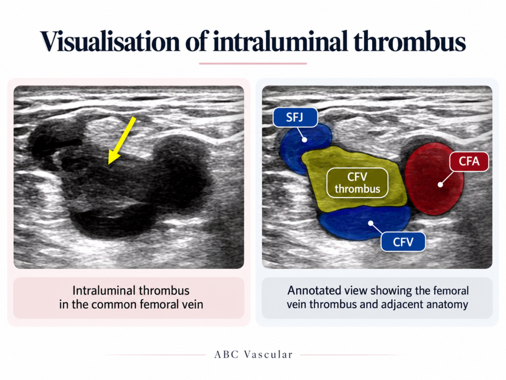
Intraluminal material may be visible on B-mode imaging, although fresh thrombus can be relatively hypoechoic and difficult to distinguish from blood. Thrombus appearance varies with composition and subsequent organisation; echogenicity alone cannot reliably establish thrombus age.
Figure 2. Visualisation of intraluminal material in the common femoral vein with annotated adjacent anatomy. Educational composite, not a standalone diagnostic example.
Colour Doppler may show a filling defect or markedly reduced flow in an involved segment. Spectral Doppler can identify associated flow abnormalities, particularly when evaluating suspected more proximal obstruction. However, an absent colour or spectral signal is not diagnostic by itself: scale, gain, wall filter, probe pressure and other machine settings can influence the display.
Acute DVT may distend the affected vein, whereas chronic post-thrombotic changes may be associated with a smaller, irregular or partially recanalised lumen. Venous calibre is an ancillary morphological feature, not a criterion to use in isolation.
Indirect Doppler findings may suggest altered venous drainage or more proximal obstruction, particularly where the affected segment is not adequately visualised. They are supportive, nonspecific signs.
Build confidence in recognising the main direct diagnostic criteria through commented scanning demonstrations, structured learning material and a CME quiz.
Explore the DVT Short Course →A focused introduction to DVT diagnosis. Full course details and eligibility are available on the course page.
Limited proximal compression ultrasound and whole-leg duplex ultrasound provide different anatomical coverage. A limited negative examination does not necessarily exclude distal disease; repeat imaging may be necessary according to the selected diagnostic pathway and the combination of clinical probability and D-dimer results.
Comprehensive duplex protocols assess proximal and calf veins with compression and targeted colour or spectral Doppler when indicated. The appropriate examination must follow local protocols, the patient’s presentation and current professional guidance. In the NICE pathway, for example, a negative proximal scan with a positive D-dimer may require a repeat scan 6-8 days later.
Venous distension and less echogenic thrombus may be associated with acute thrombosis, whereas fibrous bands, wall thickening, reduced venous diameter and recanalisation can accompany previous DVT. Individual appearances overlap, and ultrasound cannot consistently assign a precise age to thrombus.
The Society of Radiologists in Ultrasound recommends the expression chronic post-thrombotic change for residual abnormalities from previous DVT, avoiding language that could imply ongoing acute thrombosis without supporting evidence.
The ability to compress the deep vein fully remains central to ultrasound assessment for DVT. Intraluminal material, venous enlargement and Doppler abnormalities add information but must be interpreted in conjunction with proper compression technique, anatomical coverage and clinical probability.
For healthcare professional education only. Clinical decisions must follow relevant guidelines, local protocols and individual assessment.
Scientific review and final approval: Fabrizio D’Abate
Build confidence in recognising the main direct diagnostic criteria through commented scanning demonstrations, structured learning material and a CME quiz.
Explore the DVT Short Course →A focused introduction to DVT diagnosis. Full course details and eligibility are available on the course page.
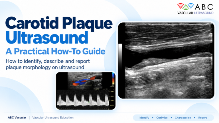
Explore the world of carotid atherosclerotic plaques with ABC Vascular’s latest eBook, “A Practical Guide on the Ultrasound Assessment of Carotid Atherosclerotic Plaques”. This guide offers healthcare professionals a comprehensive understanding of carotid plaque ultrasound assessment and its role in cardiovascular risk management.