


Explore articles, clinical insights and case studies on vascular ultrasound from ABC Vascular. Browse topics covering arterial and venous assessment, ultrasound techniques and continuing education.
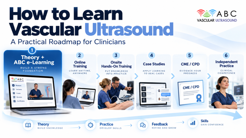
A practical staged roadmap from beginner to independent practice: core physics and machine controls, vascular anatomy and haemodynamics, probe handling and Doppler optimisation, structured protocols, carotid/peripheral arterial/venous-DVT/aortic scanning, normal-before-pathology, stenosis grading and waveform interpretation, supervised scanning with logbooks and competency assessment, reporting, common beginner mistakes, and maintaining competency via CPD/CME. Includes 2-3 simple infographics (learning pathway, competencies by stage, theory vs hands-on), a roadmap table, key takeaways, FAQs and references, ending with a restrained CTA to abcvascular.com.
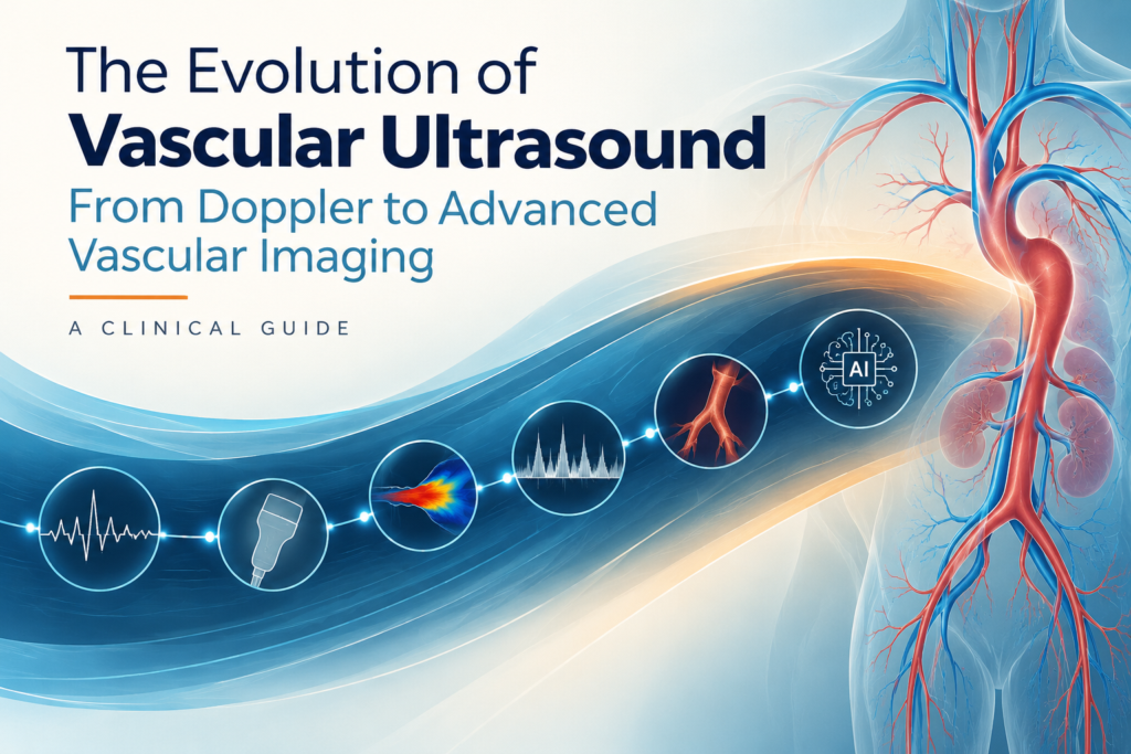
A long-form clinical history walking from continuous-wave Doppler and B-mode through duplex, spectral and colour Doppler to contrast-enhanced, 3D, elastography, microvascular and AI-assisted imaging. Uses a historical timeline, a modality-progression infographic, a clinical-applications-by-territory infographic and a past/present/future infographic, anchored by carotid, peripheral arterial, venous/DVT, aneurysm and post-intervention examples. Closes on limitations, when CTA/MRA/angiography remain necessary, and future directions.
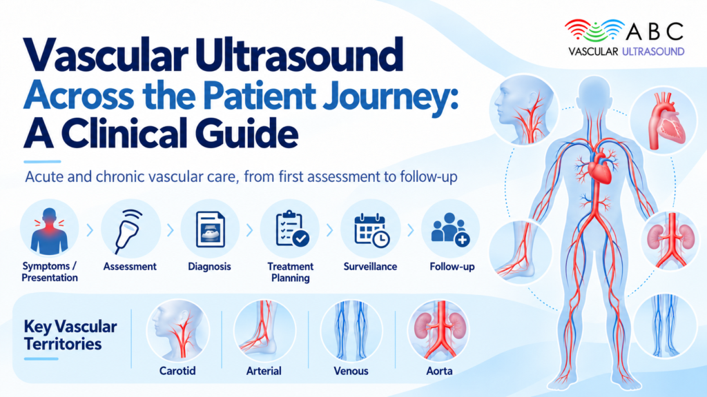
Patient-journey framework article: opens by framing five recurring clinical questions ('Is disease present?', 'How severe?', 'Haemodynamically significant?', 'Progression?', 'Has treatment worked?'), then works through carotid, peripheral arterial, venous, aortic and other territories (renal, mesenteric, upper limb) with practical examples at each journey stage. Covers advantages, limitations and operator dependence, when CTA/MRA/angiography is still needed, key takeaways, FAQs, and a restrained CTA to abcvascular.com.
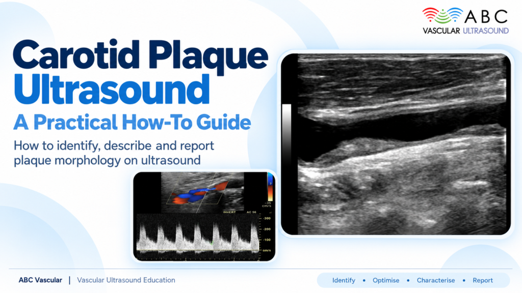
Why plaque characterisation matters: moving beyond percentage stenosis
Step-by-step protocol: grayscale, colour and Doppler settings for assessing plaque
Reporting plaque morphology: echogenicity, surface irregularity, ulceration and what to document

Explore how carotid stenosis grading and plaque characterisation influence carotid management. This article compares the two ultrasound assessment approaches and their impact on stroke risk stratification, surveillance intervals, medical therapy and referral decisions. Learn how combining stenosis measurements with plaque morphology can support more informed clinical decision-making.

A step-by-step how-to that defines IMT and plaque, contrasts their measurement rules and thresholds, walks through practical scanning examples.

Carotid ultrasound and cardiovascular risk assessment provide complementary insights into cardiovascular health. While risk scores such as SCORE2 estimate the probability of future cardiovascular events, carotid ultrasound can identify existing subclinical atherosclerosis, including carotid plaque.
This article explores how carotid imaging can refine cardiovascular risk stratification in selected patients, when ultrasound findings may influence preventive strategies, and why imaging results should always be interpreted alongside clinical risk factors and established guidelines.

When should carotid ultrasound be considered in an asymptomatic patient? Explore the role of selective screening, the ESVS 2023 guidance, and how ultrasound findings can inform cardiovascular prevention.

Vertebral artery hypoplasia on ultrasound is a common anatomical finding that can be mistaken for vertebral artery stenosis. Understanding the differences is essential for accurate vascular ultrasound interpretation and reporting.
This practical guide answers seven frequently asked clinical questions, covering vertebral artery diameter criteria, colour and spectral Doppler findings, differentiation from stenosis, right versus left anatomical variants and potential implications for posterior circulation assessment. Learn how to interpret vertebral artery hypoplasia in its clinical context and communicate findings clearly in ultrasound reports.

Internal carotid artery stenosis grading requires more than a single peak systolic velocity (PSV) measurement. Accurate carotid ultrasound assessment combines multiple Doppler parameters, anatomical findings and clinical context.
This practical guide compares NASCET and ECST grading methods, explains the role of PSV, end-diastolic velocity (EDV) and the ICA/CCA ratio, and explores common diagnostic pitfalls, including near-occlusion, contralateral carotid disease and atrial fibrillation. Learn why a multi-parameter approach supports more reliable stenosis classification and clearer vascular ultrasound reporting.