
A practical staged roadmap from beginner to independent practice: core physics and machine controls, vascular anatomy and haemodynamics, probe handling and Doppler optimisation, structured protocols, carotid/peripheral arterial/venous-DVT/aortic scanning, normal-before-pathology, stenosis grading and waveform interpretation, supervised scanning with logbooks and competency assessment, reporting, common beginner mistakes, and maintaining competency via CPD/CME. Includes 2-3 simple infographics (learning pathway, competencies by stage, theory vs hands-on), a roadmap table, key takeaways, FAQs and references, ending with a restrained CTA to abcvascular.com.
 and
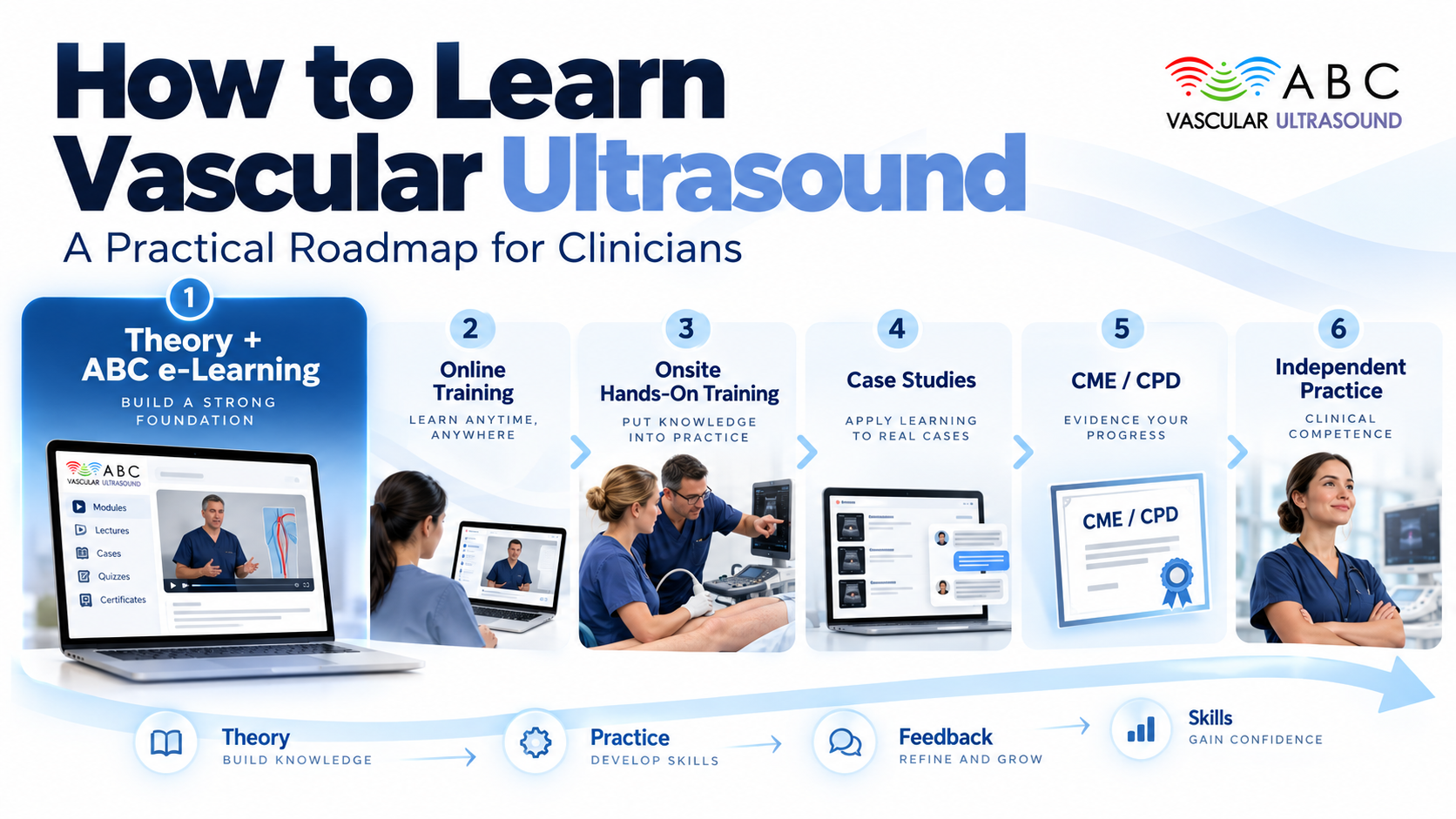
Vascular ultrasound is one of the most operator-dependent investigations in medicine. The same artery can look normal in one pair of hands and dangerously diseased in another, because the answer lives in probe angle, Doppler settings and pattern recognition as much as in the machine. For doctors, sonographers, vascular scientists, radiographers and nurses who want to learn vascular ultrasound properly, that reality shapes everything: you need a structured pathway that builds knowledge and hands-on skill together, in the right order.
Accredited e-learning is the fastest way to absorb the theory – physics, anatomy, protocols, grading criteria – and it is genuinely valuable for vascular ultrasound training. But knowledge is only half of competence. Scanning is a psychomotor skill: you learn where to place a linear probe, how to rock it to a Doppler angle of 60 degrees, and how to tell real flow from artefact by repetition, under supervision, with feedback.
Think of it like learning to drive. The highway code can be memorised in a weekend, but no one passes a driving test on theory alone. Vascular ultrasound competency works the same way: theory tells you what to look for, supervised scanning teaches your hands to find it reliably.
The learning pathway:
Beginner (theory + basics) -> Intermediate (protocols + normal findings) -> Advanced (interpretation + reporting) -> Independent Practice (consistency + CPD)
Each stage has a defined knowledge base and a defined practical milestone. Do not skip stages – most scanning errors trace back to a beginner trying to grade a stenosis before they can reliably find the vessel.
You cannot optimise an image you do not understand. Start with the essentials: how frequency trades penetration against resolution, what gain, time-gain compensation, depth and focal zone actually change, and why a 5-7 MHz linear probe suits superficial arteries while a curved probe is needed for the aorta.
Practical example: turn the depth up too far during a carotid scan and the artery shrinks to a thin stripe, tempting you to overestimate stenosis. Bring the depth down and the same vessel becomes easy to measure.
Learn the arterial and venous trees you will scan: common, internal and external carotid arteries; the vertebral arteries; the upper and lower limb arteries; the deep and superficial venous systems; and the abdominal aorta. Pair anatomy with haemodynamics – laminar flow, pulsatility, resistance, and how a stenosis converts a low-resistance waveform into a high-resistance one downstream.
This is where duplex ultrasound training becomes physical. Practise holding the probe with a light, stable grip, using the patient’s neck or limb as a fulcrum. Learn to set colour Doppler scale just above the expected peak velocity, keep a Doppler angle of 60 degrees or less, and use a small sample volume placed in the centre of the lumen.
Follow a repeatable protocol every time. For a carotid ultrasound training session, for example, scan the common carotid, then the bulb, then internal and external carotid arteries in both B-mode and colour, then obtain spectral waveforms at defined points. A fixed sequence reduces missed segments and makes your findings reproducible.
Spend deliberate hours learning what normal looks like – smooth intima, triphasic peripheral waveforms, a phasic venous signal that varies with respiration. Only once normal is familiar can you spot the abnormal with confidence. Many beginners over-call disease simply because they have never studied enough healthy vessels.
Move from pictures to numbers. Practise measuring peak systolic velocity, end-diastolic velocity and the internal carotid/common carotid ratio, and correlate them with the recognised velocity criteria. Learn to read waveform shape: loss of the triphasic pattern in a leg artery suggests significant upstream disease, while a monophasic, high-velocity signal in the internal carotid points to a tight stenosis.
Practical example: a peak systolic velocity above the accepted threshold in the internal carotid artery, combined with post-stenotic turbulence and a raised ratio, supports a high-grade stenosis – a judgement you should only make once your measurement technique is stable.
Go beyond the degree of narrowing. Describe plaque as smooth or irregular, homogeneous or echolucent, calcified or soft. Echolucent, irregular plaque carries different clinical weight from a uniform calcified plaque, and structured reporting of these features is a genuine marker of advanced vascular ultrasound competency.
A scan is only as useful as the report it produces. Learn to write structured reports that state the vessels examined, the velocities measured, the conclusion in plain language, and the clinical question answered. Link findings to management – for example, a haemodynamically significant carotid stenosis is a referral, not just a number.
Formal vascular ultrasound training requires supervised scanning with documented case volumes. Keep a logbook recording each study – the vessels, the findings, the supervising clinician – and ask for structured feedback after every session. Competency is normally signed off against a checklist: probe handling, image quality, measurement accuracy and report clarity. Aim for graded independence: observe, then scan with direct help, then scan with a supervisor available, then scan alone with audit.
Scanning too fast, before locating a stable reference point Using too much probe pressure, which compresses veins and hides thrombus Ignoring the Doppler angle and so mis-reading velocities Confusing normal anatomical variants with pathology Trusting a single velocity measurement instead of corroborating with the waveform and the B-mode image Writing reports that describe findings but never answer the clinical question
Theory and hands-on are not alternatives – they are two tracks that must advance together:
Theory track: physics – anatomy – protocols – grading criteria – reporting Hands-on track: probe skills – image optimisation – normal variants – supervised cases – independent scanning
Coursework can carry most of the theory track quickly. The hands-on track only grows with supervised scanning time, which is why online learning alone never produces a competent operator.
Competence is not a certificate you frame and forget. Accuracy decays with low case volume, so protect regular scanning time and keep a spread of studies across carotid, peripheral arterial, venous/DVT and aortic work. Maintain CPD/CME through accredited vascular ultrasound courses, audit your reports against outcomes, and revisit the fundamentals when new guidelines appear.
Competence requires both theoretical knowledge and supervised hands-on scanning – neither alone is sufficient Follow a staged roadmap: Beginner, Intermediate, Advanced, Independent Practice Learn normal findings before you attempt to grade pathology Measure velocities with a correct Doppler angle and corroborate every number with the waveform and B-mode image Document supervised scans in a logbook and seek structured feedback Protect case volume and CPD/CME to keep your accuracy after you qualify
No. Accredited online courses are excellent for the theory – physics, anatomy, protocols and grading criteria – but vascular ultrasound competency depends on supervised hands-on scanning with feedback. Use online learning to build knowledge, then transfer it to supervised clinical practice.
It varies with prior experience and caseload. A focused beginner can cover the theory in weeks, but reaching independent practice typically takes months of supervised scanning and a documented logbook of cases across carotid, peripheral arterial, venous and aortic studies.
A structured vascular ultrasound course gives you the framework – a defined curriculum, protocols and a competency checklist – so you do not learn bad habits. Ideally choose accredited e-learning in duplex ultrasound training and Doppler ultrasound training, then pair it with supervised clinical scanning.
Society for Vascular Surgery and international consensus guidelines on carotid and peripheral arterial velocity criteria EFSUMB and UEMS guidance on vascular ultrasound training and competency standards Published duplex criteria for the diagnosis of lower limb deep vein thrombosis and peripheral arterial disease Accreditation and CME/CPD requirements for vascular ultrasound practice
Ready to build the theory side of your roadmap? ABC Vascular’s accredited online courses and the 2026 Vascular Ultrasound Book give clinicians a structured, self-paced foundation in vascular ultrasound – then take it to your scanner. Start at abcvascular.com.