
A step-by-step how-to that defines IMT and plaque, contrasts their measurement rules and thresholds, walks through practical scanning examples, and closes with a CTA to the Carotid and Vertebral Arteries master course.
 and
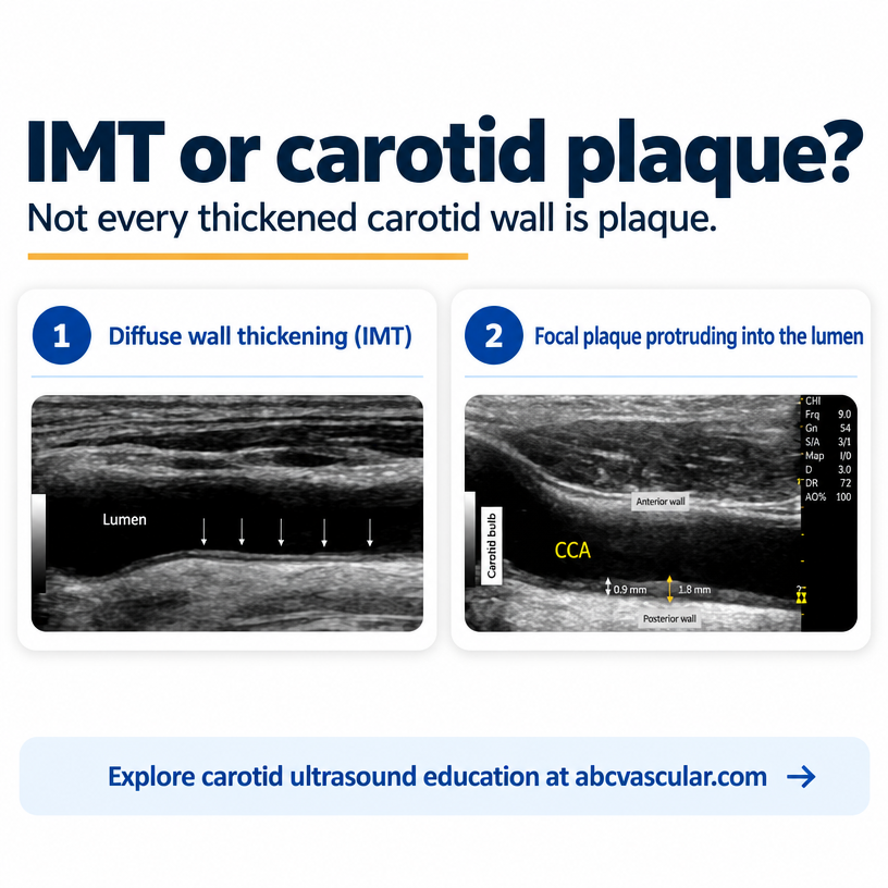
Few measurements in vascular ultrasound are confused as often as intima-media thickness (IMT) and plaque. On a greyscale B-mode image, both appear as thickening of the arterial wall, and at a glance the two can look almost identical. Yet they describe different biological processes, follow different measurement rules, and carry different implications for the patient lying in front of you.
Getting the call right changes whether a scan is reported as an age-related finding, an early marker of systemic atherosclerosis, or a focal lesion that needs grading and follow-up. This guide walks through the distinction step by step, with the practical examples you meet on real scanning lists – and shows how the two are assessed side by side in the accredited Carotid and Vertebral Arteries master course.
IMT is a diffuse, measured property of the arterial wall. Plaque is a focal lesion that encroaches on the lumen. One is a number in millimetres describing the wall as a whole; the other is a discrete structure you find, characterise and grade.
Reporting one as the other is not harmless shorthand. Call a focal plaque ‘just thickening’ and you risk leaving lumen encroachment, surface characteristics and local stroke risk unrecorded. Label a diffusely thickened wall ‘plaque’ and you may over-diagnose disease, trigger unnecessary anxiety, and dilute the meaning of the word across your reports. Clear, consistent language is what your referring clinicians depend on.
Intima-media thickness is the combined width of the intima and the media. On B-mode ultrasound you measure it from the leading edge of the intima-lumen interface to the leading edge of the media-adventitia interface, taking care not to include the adventitia itself.
In practice, IMT is measured on the far wall of the common carotid artery, in a strictly plaque-free segment, usually over a 1 to 2 cm length proximal to the flow divider. It is a continuous variable: every patient has a value, and that value is compared with age- and sex-referenced norms.
The thresholds that guide reporting are widely agreed. A value below 0.9 mm is generally regarded as normal in adults. A value between 0.9 and 1.5 mm represents increased or evident thickening. A wall thickness above 1.5 mm, measured from the media-adventitia interface to the intima-lumen interface, is itself treated as plaque under the Mannheim consensus – which is precisely where the two concepts begin to overlap.
A carotid plaque is a focal structure that encroaches into the arterial lumen. The Mannheim consensus formalises this definition: a plaque is present when a focal structure encroaches at least 0.5 mm into the lumen, or by at least 50 per cent of the surrounding IMT value, or when the wall thickness exceeds 1.5 mm as measured from the media-adventitia interface to the intima-lumen interface.
The word doing the work here is focal. Plaque is a local, asymmetric lesion, not a diffuse property of the vessel. That is why a focal thickening that bulges into the lumen is a plaque even if the surrounding wall is thin, and why a uniformly thickened wall without any focal encroachment is increased IMT rather than plaque.
Once you have called a plaque, the job is only half done. A plaque is characterised, not just detected: greatest thickness, cross-sectional area, echogenicity (echolucent, mixed or calcified), texture (homogeneous or heterogeneous), surface (smooth, irregular or ulcerated) and mobility. Echolucent, heterogeneous and ulcerated plaques are the features most consistently linked to increased embolic risk, which is why the description matters as much as the diagnosis.
IMT is diffuse; plaque is focal. IMT is measured in a defined plaque-free segment; plaque is measured at the lesion itself. IMT is a continuous value in millimetres; plaque is a categorical finding with a characterisation attached. IMT is primarily a marker of systemic cardiovascular risk; plaque is a focal atherosclerotic lesion that can carry local stroke risk through its surface and composition.
A simple way to hold the distinction in mind: IMT tells you about the arterial wall as a whole, while plaque tells you about one specific place in the artery. The two can coexist, and often do – but they answer different clinical questions, so they should always be reported separately.
Set the patient supine with the head rotated slightly away from the side being scanned, and use a high-frequency linear array transducer – typically 7 to 12 MHz for the common carotid artery.
Image the common carotid artery in longitudinal B-mode, adjusting the focal zone and time-gain compensation so the intima-lumen and media-adventitia interfaces are crisp and the beam strikes the wall perpendicularly. A beam that is not perpendicular thickens the apparent wall and inflates your value.
Choose a plaque-free segment 1 to 2 cm proximal to the carotid bulb. Avoid the bulb and the flow divider, where the wall is normally thicker and the geometry is less reproducible.
Measure the far wall only. The near wall is systematically overestimated because of refraction and beam-width artefacts, so it is not used for routine IMT measurement.
Take several measurements across the segment and report the mean, and use semi-automated edge-detection software where it is available to reduce operator variability. Consistency of technique matters more than any single reading – the same segment, the same plane, the same landmarks every time.
Scan the whole accessible extracranial course in both longitudinal and transverse planes. Plaques are frequently asymmetric, so a single longitudinal sweep can miss a lesion that a transverse view displays clearly.
Look for a focal, asymmetric thickening that encroaches on the lumen, and confirm any candidate lesion in two orthogonal planes before you commit to the diagnosis. This step alone removes most false positives, because diffuse thickening can imitate focality in one plane.
Measure maximal plaque thickness from the media-adventitia interface to the intima-lumen interface, and record the cross-sectional area where your protocol calls for it. Then describe the plaque: echogenicity, homogeneity, surface regularity, calcification and any mobile component.
Document each plaque with representative images and, where possible, a diagram. A structured description is what allows a colleague – or the same sonographer at the next scan – to track how the lesion changes over time.
A 62-year-old man with well-controlled hypertension attends for a carotid assessment. His common carotid far-wall IMT measures 1.1 mm diffusely on both sides, with smooth interfaces and no focal encroachment anywhere along the common carotid artery, bulb or internal carotid artery origin.
The correct report is increased intima-media thickness, no plaque identified. This is a systemic risk marker. It supports attention to blood-pressure control, lipids and lifestyle, and it may reclassify the patient’s cardiovascular risk – but it is not a focal plaque, and it is not graded as a stenosis.
If you were tempted to write ‘plaque’ because 1.1 mm feels thick, this example shows the cost of that shorthand: the referring clinician loses the distinction between a diffuse marker and a focal lesion, and future scans lose their baseline.
A 58-year-old woman who smokes has a common carotid IMT of 0.7 mm – squarely normal. But at the right internal carotid artery origin there is a 2.4 mm echolucent plaque that encroaches clearly into the lumen, with an irregular surface.
This is the opposite trap: a normal IMT with a significant focal plaque. The IMT value tells you nothing reassuring here, because the disease is focal. The report should state normal IMT and a focal right ICA origin plaque, then characterise it – echolucent, irregular, 2.4 mm thick – and grade the stenosis on Doppler criteria.
This example is a useful reminder that a normal IMT never excludes plaque. The two findings are independent, and either can be present without the other.
A 70-year-old man with type 2 diabetes has both: a diffusely thickened common carotid wall at 1.3 mm, and a calcified 3.0 mm plaque at the left carotid bulb with acoustic shadowing.
Here the two findings coexist, and the correct approach is to report them separately rather than collapsing them into one. State the increased IMT as a systemic marker, then describe the bulb plaque in full – thickness, echogenicity, calcification and surface. Avoid double-counting the same millimetres in both the IMT value and the plaque thickness, and keep your IMT measurement strictly within the plaque-free segment.
Measuring IMT inside a plaque segment is the most common error; it inflates the value and confuses the two concepts. Including the adventitia in the measurement is the second, and it silently overestimates thickness. Measuring the near wall rather than the far wall is the third, for the artefact reasons already described. Measuring at the bulb or flow divider is the fourth, because the wall is normally thicker there. And calling any thickness above 1.5 mm plaque without checking whether the thickening is focal is the fifth.
Each of these is a small technique slip, but together they are the reason two sonographers can look at the same artery and report different things. Standardising your landmark, plane and gain settings fixes most of it.
Increased IMT indicates that atherosclerosis is developing across the vascular system. It is a risk marker that informs global cardiovascular risk assessment and supports decisions about medical therapy and lifestyle change. It is not, by itself, a focal lesion and it is not graded.
Plaque indicates focal atherosclerotic disease and adds local information: lumen encroachment, surface characteristics and composition. Echolucent, heterogeneous and ulcerated plaques carry the greatest embolic concern, while heavily calcified plaques are often more stable. Detecting plaque can reclassify a patient into a higher-risk category and change the intensity of follow-up.
Read together, the two findings give a fuller picture: the IMT describes the background, the plaque describes the lesion. Reporting both, in their own terms, is what makes a carotid scan genuinely useful to the clinician acting on it.
Reading about the IMT-versus-plaque distinction is one thing; making the call confidently, on a moving image, with the gain settings you actually have, is another. That practical confidence comes from repetition on real cases, and it is exactly what the Carotid Ultrasound Course and Vertebral Arteries is built to deliver.
The master course covers the full extracranial assessment – basic carotid Doppler principles, anatomy, step-by-step scanning protocols, and the ultrasound criteria for detecting, characterising and grading carotid, vertebral and subclavian disease – through commented video lectures, e-booklets, real case studies and interactive quizzes. It is accredited and awards 20 AMA PRA Category 1 Credits, and you can start with the first lessons for free before you commit.
If you want your IMT measurements to be reproducible and your plaque descriptions to be consistent, work through the accredited Carotid and Vertebral Arteries master course and practise on the case library until the distinction becomes second nature. Learning is not a destination, it is a journey – and this is the step that sharpens your daily reporting.
IMT is a diffuse, continuous measurement of the intima and media in a plaque-free segment, usually the far wall of the common carotid artery, with values below 0.9 mm normal, 0.9 to 1.5 mm increased, and above 1.5 mm treated as plaque under the Mannheim consensus. Plaque is a focal, asymmetric lesion that encroaches on the lumen – defined as at least 0.5 mm encroachment, or 50 per cent of the surrounding IMT, or a thickness above 1.5 mm – and it must be characterised, not just detected. Measure IMT away from plaque, report the two findings separately, and never let a reassuring IMT value talk you out of fully assessing a focal lesion.