Common femoral artery occlusion and retrograde flow in the profunda femoris artery: Doppler waveforms scenario

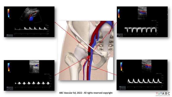
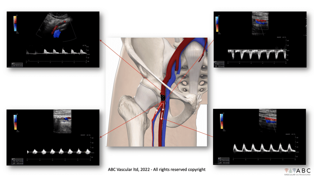
Case study description: In this case a set of pulse wave Doppler waveforms sampled in a patient with an occluded common femoral artery are shown. The Doppler waveforms are sampled in the distal external iliac artery proximally to the occluded common femoral artery (CFA), distal to the occluded common femoral artery, within the proximal profunda femoris artery (PFA) and in the proximal superficial femoral artery (SFA).
Video length: 0 mins
Audio: No

In presence of an occlusion of the common femoral artery, the most common collateral pattern for supplying blood flow to the leg is via retrograde flow from the profunda femoris artery into the superficial femoral artery. Retrograde flow within the PFA enters the SFA passing via the distal patent lumen of the CFA and femoral bifurcation.
Retrograde flow from the PFA to the SFA can be detected both by using colour flow Doppler and pulsed wave Doppler.
Colour flow Doppler would demonstrate the PFA and the SFA presenting two different colour codes (in this case red in the SFA – flow toward the transducer sliding down from the inguinal canal toward the popliteal fossa and blue in the PFA – flow away from the transducer according to the colours seen in the lateral colour scale bar).
Blood flow direction can be confirmed and better defined using pulsed wave Doppler. As shown in this case, the Doppler waveform recorded within the PFA presents retrograde flow as demonstrated by the presence of flow below the baseline. The SFA presents antegrade flow as the Doppler waveform is seen above the baseline. The SFA waveform analysis shows a monophasic flow with low velocities as per proximal obstruction. Flow within the distal external iliac artery demonstrate a typical pre-occlusive monophasic high resistance flow.
Such collateral path is possible when the occlusion of the CFA does not involve the femoral bifurcation and/or the distal segment of the CFA, as demonstrated in this case where the distal CFA and proximal superficial and profunda femoris artery are patent. Doppler waveform sampled in the distal patent CFA are generally monophasic with low velocity waveforms as per the presence of a proximal occlusion, while Doppler waveforms sampled proximally to the occluded CFA, in the distal external iliac arteries, are generally monophasic high resistance type with low peak systolic velocities, a waveform typically describing a pre-occlusive flow state.
Take home messages: in presence of an occluded CFA, flow in the profunda femoris artery is generally retrograde as it acts as the main collateral pathway to fill the SFA. Such pattern is possible when the femoral bifurcation is patent.


