Deep Vein Thrombosis in Intravenous Drug Users

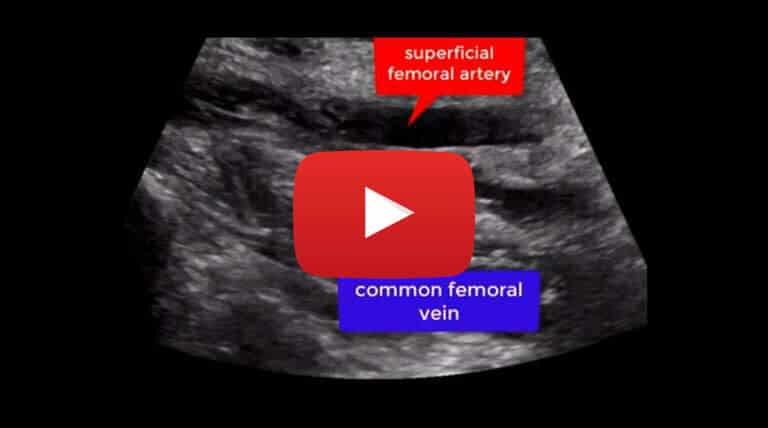
Case study description: Short video on DVT in intravenous drug users
Video length: 3:45 mins
Audio: Yes, with voice-over explanation
Addiction to intravenously administered drugs is a serious epidemiological problem causing several health complications. Among these, deep vein thrombosis (DVT) is one of the most important. Up to one in five DVTs occur in intravenous drug abusers (IVDAs) and almost half of the DVTs in under forties have been related to intravenous drug abuse.
Other vascular and non-vascular complications include: seroma, venous and arterial pseudoaneurysm, haematoma, infection, scar tissue, infection.
This short video shows the most common ultrasound features of DVT in IVDAs patients. Ultrasound operators should be familiar with the typical DVT ultrasound findings in IVDAs as they differ from common DVT ultrasound appearance in the general population.
The most common ultrasound findings encountered when scanning DVT in IVDAs are the following:
- Proximal femoral veins in the area of a puncture are often the site of DVT
- The common femoral vein can result stenosed or focally collapsed or atretic
- When visible, the calibre of vein used for the injection can appear small or thickened
- Iliac veins can be partially or completely thrombosed or with scarring thrombus
- An acoustic shadow is often visible in the groin at the level of puncture
- Presence of venous collateralisation upon occlusion of the common femoral vein
The groin is the most common site used by IVDAs for self-injecting, thus increasing the risk of developing ilio-femoral DVTs which are the most common type of DVTs in these patients.
Often the common femoral vein is damaged due to repetitive injections and sometimes can appear of narrow calibre with evidence of venous wall thickening or peri-venous walls oedema (appearing as the halo sign in vasculitis).
Stenosis of the common femoral vein is common and so are collapsed segments of this vessel as a consequence of the repetitive injections.
Scarring around injection sites is often present and can generate acoustic shadow at the injection site. The scarring is caused by chronic indurative tissue at the site of injection due to repetitive injections.
Venous collaterals are often visible at the level of the common femoral vein as a result of a long lasting occlusion, stenosis or collapse of this vessel. These collaterals can also be the result of thrombotic occlusion of the great saphenous vein and other tributary veins emptying into the saphenofemoral junction and can manifest in a later stage as varicose veins.
These tortuous varicose veins may extend over a large area of the thigh and can occasionally extend over the lower anterior abdominal and pelvic wall to the genitalia.
The use of Color Doppler Flow is mandatory to better depict the course and flow direction within venous collaterals.
We suggest the master course on venous ultrasound to learn more.


