
Comparison-shaped article contrasting probability-based risk tools with carotid ultrasound's ability to detect subclinical atherosclerosis and refine risk in selected patients, grounded in ESC 2024 and ESC/EAS 2025, with SEO headings, key takeaways, references and one CTA.
 and
Cardiovascular risk scores and carotid ultrasound answer two fundamentally different questions. A risk score estimates the probability that a person will have a cardiovascular event over a defined period, using population-derived variables. Carotid ultrasound examines the artery itself and can show whether atherosclerosis is already present. Recognising that distinction is the key to using imaging intelligently: it clarifies when a scan genuinely refines risk assessment, and when it adds cost and uncertainty without changing management.
For sonographers, physicians and clinicians, the practical question is not whether imaging is ‘better’ than a risk score. It is which patients benefit from looking directly at the arterial wall, and how to interpret what the scan shows.
Risk tools estimate probability from population data; they do not confirm whether an individual already has atherosclerosis.
Carotid ultrasound can detect subclinical atherosclerosis, including plaque and increased intima-media thickness, before symptoms appear.
Imaging is most informative in selected patients, especially when calculated risk is borderline or when the decision to intensify prevention is finely balanced.
Guideline frameworks including ESVS 2023, ESC 2024 and ESC/EAS 2025 support targeted rather than indiscriminate use of vascular imaging.
An ultrasound finding informs clinical judgement; it does not by itself mandate a particular drug or procedure.
Tools such as SCORE2 and SCORE2-OP estimate the ten-year probability of a first cardiovascular event in apparently healthy adults. They draw on variables that are quick to collect – age, sex, smoking status, systolic blood pressure and cholesterol – and compress them into a single figure.
That figure is a probability, not a diagnosis. A ten-year risk of 12% means that, in a comparable group of one hundred people, roughly twelve might be expected to have an event over a decade. It does not reveal whether the person in front of you already has plaque, how much, or in which artery.
This is the central limitation of risk scoring. Most people who experience a cardiovascular event fall into intermediate-risk bands, yet most people in those bands never have an event. A tool designed to rank a population cannot, on its own, resolve the individual question of whether disease is present.
Risk scores are also sensitive to how they are used. Reclassifying a borderline score can tip a management decision, which is exactly why clinicians look for additional information. Traditional risk factors do not capture every important influence, and some people with modest scores harbour substantial disease while others with higher scores do not.
This is where direct imaging of the arterial tree becomes attractive. If a score tells us the odds, imaging tells us something about the arteries themselves.
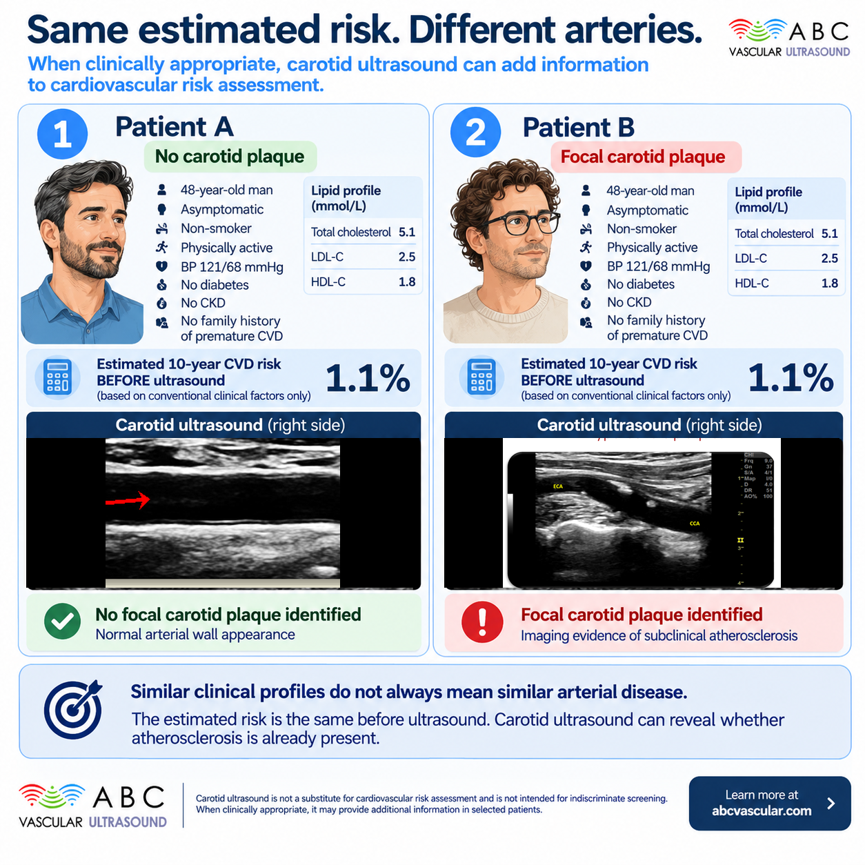
Carotid ultrasound interrogates the arterial wall directly. It can identify two things a risk score cannot: focal atherosclerotic plaque, and increased intima-media thickness. Both are markers of subclinical atherosclerosis – disease that is present but has not yet produced symptoms.
When plaque is present, the clinician is no longer reasoning about probability alone. There is direct evidence that the atherosclerotic process has begun in that patient. In selected individuals, this can move a risk estimate upward and support a more assertive approach to prevention.
It is important to be precise about what this means. Detecting plaque is an imaging finding, not a treatment decision. The ESVS 2023, ESC 2024 and ESC/EAS 2025 frameworks treat carotid plaque as a risk modifier – information that refines a risk estimate – rather than an automatic trigger for a specific intervention. The finding is one input into a wider clinical assessment that also weighs blood pressure, lipids, glucose, lifestyle and patient preference.
Two patients can both have plaque yet warrant different conversations. Plaque burden, its acoustic characteristics and the overall clinical picture all shape what the finding means for that individual.
A plaque detected in a patient with borderline risk and few other concerns may prompt a discussion about earlier or more intensive risk-factor control. The same finding in a patient already committed to treatment may change little. The scan refines judgement; it does not replace it.
For this reason, the ultrasound report matters as much as the scan. Clear documentation of plaque presence, location, extent and acoustic appearance gives the treating clinician something they can actually use.
A carotid ultrasound report does not arrive in isolation. It joins a clinical picture, and its meaning depends on that picture.
Consider a patient with well-controlled blood pressure, no smoking history and a borderline risk score. A normal scan is reassuring and supports continuing current management. A plaque finding may prompt a conversation about earlier or more intensive lipid-lowering.
Now consider a patient with a strong family history, hypertension and a borderline score. The same plaque finding carries more weight because it aligns with a suspicion already present.
In both cases the ultrasound is valuable not because it issues an instruction, but because it reduces uncertainty in a way that lets clinician and patient decide together.
If carotid ultrasound reliably reveals disease, why is it not offered to everyone? Because the value of any test depends on who receives it.
In a population with very low pre-test probability, most scans will be normal, and a normal result rarely changes management. In a population already at high risk – for example, someone with established cardiovascular disease or diabetes with target-organ damage – the decision to intensify prevention is usually clear without imaging. The test adds most where it resolves genuine uncertainty.
That middle ground is where selective imaging earns its place. A patient whose calculated risk sits near a decision threshold, or whose risk factors are numerous but whose overall picture is ambiguous, may benefit from knowing whether plaque is present. One well-chosen piece of information can tip a finely balanced decision.
This is the essence of selectivity. It is the opposite of indiscriminate screening, and it respects both the limits of the technology and the principle that tests should change what we do.
For clinicians weighing whether to image, a few questions keep the decision disciplined:
Is the calculated risk borderline, or discordant with the clinical picture?
Would the presence or absence of plaque change management?
Are there multiple vascular risk factors that together raise suspicion?
Is the patient able to undergo and understand the test, and will the result inform a shared decision?
If the answer to these questions is consistently ‘no’, imaging is unlikely to add value. If the finding could reasonably change what you do next, it has earned its place.
When imaging is used, it should be done to a standard. That means a reproducible acquisition protocol, consistent terminology, and reporting that a non-specialist colleague can act on. It also means knowing the difference between a finding that changes management and a finding that merely confirms what was already suspected.
Carotid assessment is a skill. Interrogating the wall, recognising plaque and describing it reproducibly are learned abilities that improve with structured practice.
Several ideas recur in day-to-day practice and are worth correcting.
Plaque equals a mandate for drugs. This is not how the guidance reads. Plaque is a risk modifier; treatment decisions remain clinical and individual.
A normal carotid scan rules out cardiovascular disease. It does not. Atherosclerosis is a systemic process, and a single arterial bed is only a window onto it.
A high risk score means imaging is unnecessary. Not always. The score reflects probability; the scan reflects disease presence, and the two can disagree in clinically meaningful ways.
Risk scores and carotid ultrasound are complementary. Scores provide a population-grounded probability; ultrasound provides an individual assessment of disease presence. Used together, and used selectively, they can refine cardiovascular risk assessment in exactly the patients where refinement matters.
The evidence-based position is measured: imaging is a tool for sharpening risk assessment in the right patient, not a screening ritual for everyone. That is where its clinical value concentrates.
For professionals who want to build confidence in acquiring and interpreting carotid studies, structured, accredited training provides the foundation.
Learn more at abcvascular.com.
European Society for Vascular Surgery (ESVS). 2023 Clinical Practice Guidelines on the Management of Atherosclerotic Carotid and Vertebral Artery Disease. European Journal of Vascular and Endovascular Surgery.
European Society of Cardiology (ESC). 2024 ESC Clinical Practice Guidelines. European Heart Journal.
European Society of Cardiology / European Atherosclerosis Society (ESC/EAS). 2025 Guidelines for the management of dyslipidaemias. European Heart Journal.