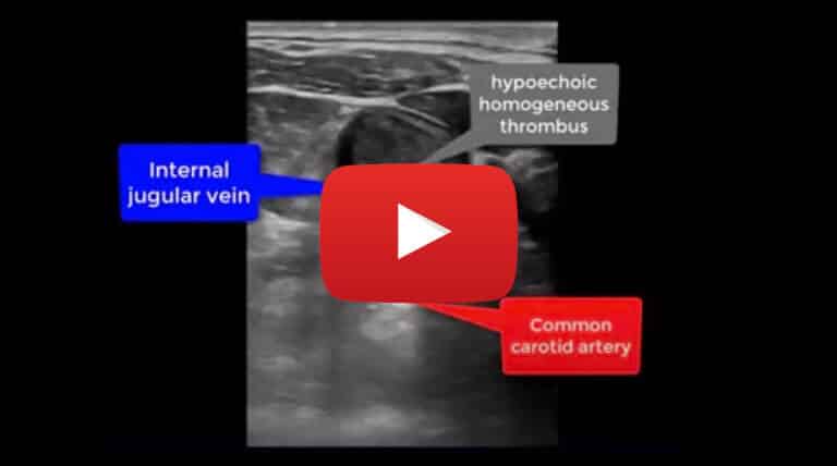
Acute Deep Vein Thrombosis of the Internal Jugular Vein: B-Mode Imaging

Case study description: Short video showing acute DVT of the IJV
Video length: 33 secs
Audio: No audio
This video shows a hypoechoic thrombus attached to the anterior wall of the internal jugular vein with the remaining part of the thrombus fluctuating within the IJV lumen. The common carotid artery is shown posteriorly to the IJV.
The common carotid artery is noted posteriorly to the IJV both in transverse and longitudinal view. This is one of the main anatomical landmarks used to identify the IJV.


