A Thrombosed Common Femoral Artery Pseudoaneurysm: Ultrasound Features

Case study description: Short video showing the features of a thrombosed common femoral artery pseudoaneurysm
Video length: 40 secs
Audio: No audio
A large perfused pseudoaneurysm of the common femoral artery (CFA) was detected with ultrasound in a patient that had developed groin pulsatile swelling after a TAVI procedure.
The pseudoaneurysm was treated with thrombin injection as a small pseudoaneurysm neck (<5 mm) was present.
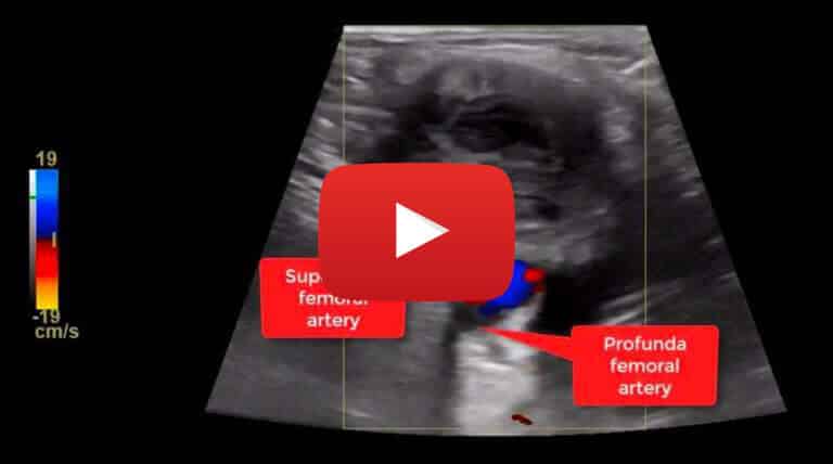
The video shows the B-mode and Color Doppler Flow ultrasound features of the pseudoaneurysm after being treated with thrombin injection.
On Color Doppler Flow no evidence of perfused areas were noted within the pseudoaneurysm and no connection with the common femoral artery was present.
Low Color Doppler Flow scale was used to detect low velocity flow. The colour flow scale should be progressively reduced in order to detect low flow, taking into consideration that colour bleeding artefact can occur and can be misleading for the presence of flow within the thrombosed areas.


