Iatrogenic Arterio-Venous Fistula Between the Superficial Femoral Artery and Common Femoral Vein

Case study description: Iatrogenic AVF between the superficial femoral artery and common femoral vein
Video length: 1:19 min
Audio: Yes, with voice-over explanation
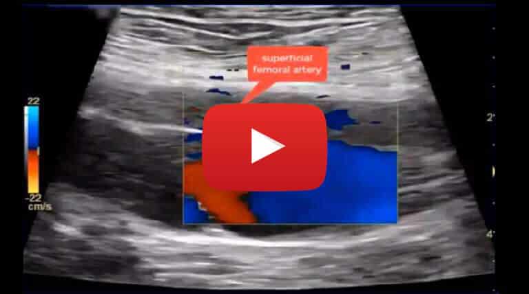
This is a case of an 81 years old male patient that underwent a cardiac ablation procedure with access via the right groin. A post procedure (3 days after) ultrasound scan was requested due to the onset of a pulsatile mass noted at the level of the right upper thigh. On the ultrasound examination, a high jet velocity arterio-venous fistula (AVF) was noted between the proximal superficial femoral artery and the distal common femoral vein. No pseudoaneurysm or haematoma were noted.
An AVF is an abnormal connection or passageway between an artery and a vein. It may be congenital, surgically created for haemodialysis treatments, or acquired due to pathologic process, such as trauma or erosion of an arterial aneurysm or iatrogenic, particularly after percutaneous procedures.
This is the case of an iatrogenic AVF as it appeared after the cardiac ablation procedure. The pre-op ultrasound examination of the femoral arteries was normal.
The AVF was firstly noted on Color Doppler Flow, then confirmed with the pulsed wave Doppler analysis. Color Doppler Flow allows to identify the connection between the arterial and venous vessel and understand the direction of flow. AVFs usually present with high and turbulent arterio-venous velocity flow. This can be demonstrated both with Color Doppler Flow and pulsed wave Doppler. On Color Doppler Flow the high velocity flow is seen as colour flow aliasing while on pulsed wave Doppler appears as bidirectional high velocity flow (flow above and below the zero line). Due to the communication between the artery and the vein, venous flow becomes arterialised and turbulent proximally to the AVF which is demonstrated by the presence of vortexes on Color Doppler Flow and spectral broadening on pulsed wave Doppler. The presence of the AVF tends to reduce the peripheral arterial resistance within the artery connected to the vein and, therefore, it is often possible to note a monophasic arterial waveform proximally to the AVF rather than triphasic as is normally observed in the femoral arteries. In this case the Doppler waveform recorded in the superficial femoral artery proximally to the AVF is triphasic, however, a more pronounced diastolic phase is noted. This is due to the fact that the AVF neck is of small calibre and therefore does not reduce the peripheral resistance significantly.
Take home message: To detect the presence of an AVF both Color Doppler Flow and pulsed wave Doppler should be used: Color Doppler Flow is essential to identify the connection between the two arterial and venous vessels and understand the direction of flow; then pulsed wave Doppler flow can help in confirming the presence of the AVF and obtain information on Doppler waveforms characteristics within the AVF, the vein and artery involved.


