
A long-form clinical history walking from continuous-wave Doppler and B-mode through duplex, spectral and colour Doppler to contrast-enhanced, 3D, elastography, microvascular and AI-assisted imaging. Uses a historical timeline, a modality-progression infographic, a clinical-applications-by-territory infographic and a past/present/future infographic, anchored by carotid, peripheral arterial, venous/DVT, aneurysm and post-intervention examples. Closes on limitations, when CTA/MRA/angiography remain necessary, and future directions.
 and
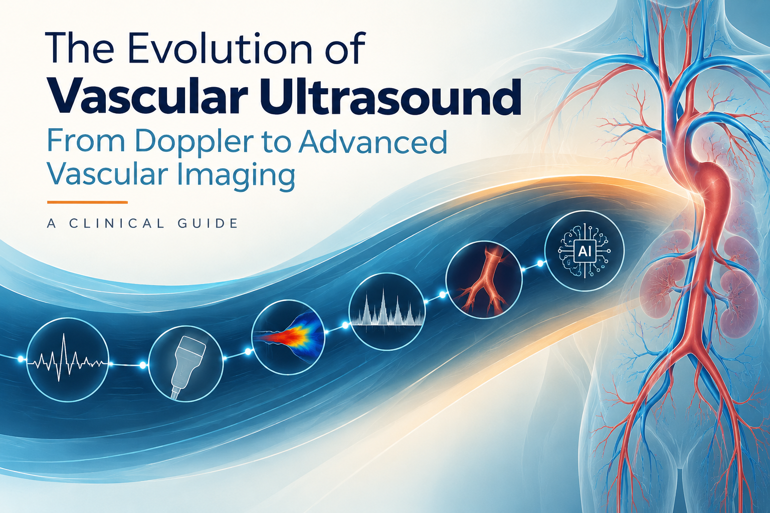
Vascular ultrasound is one of the few imaging tools that has been reinvented, rather than merely improved, over the past half century. What began as a crude way to confirm that blood was moving has become a first-line vascular imaging method that can grade stenosis, characterise plaque, quantify haemodynamics and track a patient’s response to treatment over years. For the clinician at the bedside, the important question is not simply what the images look like now, but how each technological step changed the decisions we make. This article traces that journey, from continuous-wave Doppler and basic B-mode through duplex ultrasound, spectral and colour Doppler, to contrast-enhanced ultrasound, 3D imaging, elastography, microvascular imaging and AI-assisted analysis.
Every advance in ultrasound technology answered a clinical question the previous generation could not. Detecting flow told us a vessel was patent; spectral Doppler told us how severely flow was disturbed; colour Doppler told us where that disturbance was; and newer techniques tell us whether a plaque is unstable, whether a treatment is working, and how a disease is likely to behave. The result is a shift from describing vessels to predicting outcomes.
Timeline of vascular ultrasound evolution
Each of these steps was adopted not because the images were prettier, but because they changed what a clinician could do at the point of care.
The core progression runs from simply detecting flow to measuring it, locating it, and interpreting its waveform. Continuous-wave Doppler cannot tell depth, but it is exquisitely sensitive. Pulsed (spectral) Doppler adds range resolution, so velocity can be measured at a chosen point within a vessel. Duplex ultrasound pairs that spectral trace with a real-time B-mode image, giving anatomy and haemodynamics in a single view. Colour Doppler then maps flow across the whole field, turning a single sample volume into a spatial picture. Together they moved vascular ultrasound from a presence-or-absence test to a quantitative examination.
Doppler to duplex to colour to advanced imaging
Carotid ultrasound historically answered one question: what is the degree of stenosis? Spectral Doppler criteria such as peak systolic and end-diastolic velocity and internal-to-common carotid ratios remain the backbone of grading. But colour Doppler and B-mode revealed that two plaques with the same stenosis can behave very differently. Grey-scale and echolucency assessment, surface irregularity, and later elastography and contrast-enhanced ultrasound, highlight features associated with higher stroke risk. Clinically, this supports more nuanced decisions about who benefits from intervention, which is why carotid ultrasound now informs risk stratification rather than delivering a single percentage.
In arterial ultrasound, the waveform became as important as the anatomy. A monophasic, damped spectral trace distal to a lesion tells a clinician that disease is proximal and significant, even before the lesion itself is seen. Duplex mapping of the lower limb lets an interventionist plan access and target the correct segment, while post-procedural duplex detects restenosis before symptoms return. The shift is from asking whether a blockage exists to asking where the haemodynamic bottleneck lies and how best to treat it.
Venous ultrasound transformed DVT management. Compression ultrasound, augmented by colour and spectral Doppler, distinguishes acute from chronic thrombus, assesses vein compressibility and quantifies reflux in chronic venous disease. Because the test is repeatable and radiation-free, it became the tool for both diagnosis and serial follow-up, guiding the duration of anticoagulation and identifying post-thrombotic change.
Abdominal aortic aneurysm surveillance was built on reproducible diameter measurement, and ultrasound remains the preferred modality because it is safe and repeatable. Advances now add morphology, including thrombus burden and wall characteristics, while elastography explores wall stiffness. These help clinicians interpret growth patterns and better time referral for repair rather than relying on a single diameter threshold.
After stenting, bypass or endovascular repair, duplex ultrasound is the surveillance workhorse. It detects intimal hyperplasia, graft stenosis and endoleak, and tracks patency over time. This is where the modern examination earns its value: it does not just describe disease, it measures the response to treatment and flags failure early, while it is still correctable.
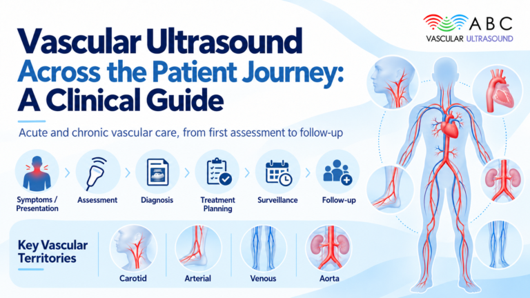
Clinical applications by vascular territory
| Territory | Primary ultrasound role | Key modalities |
|---|---|---|
| Carotid | Stenosis grading, plaque characterisation, stroke risk | Duplex, spectral and colour Doppler, CEUS, elastography |
| Peripheral arterial | Lesion localisation, haemodynamic significance, planning | Duplex mapping, spectral waveform analysis |
| Venous / DVT | Diagnosis, reflux quantification, clot characterisation | Compression, colour and spectral Doppler |
| Aneurysm | Surveillance, growth tracking, morphology | B-mode, 3D, elastography (emerging) |
| Post-intervention | Patency, restenosis, endoleak surveillance | Duplex ultrasound, colour Doppler, CEUS |
Past vs present vs future
| Past | Present | Future | |
|---|---|---|---|
| Core question | Is there flow? | What are the haemodynamics, plaque and wall doing? | What will happen to this patient, and how should care be personalised? |
| Main tools | Continuous-wave Doppler, B-mode | Duplex, colour Doppler, CEUS, 3D, elastography | AI-assisted analysis, 4D imaging, quantitative plaque analysis |
| Clinical use | Detection | Diagnosis, triage, planning, surveillance | Prediction and personalised vascular assessment |
| Limitation | No depth or detail | Operator dependence, reproducibility | Validation, standardisation, governance |
Ultrasound’s flexibility is also its weakness. Every practitioner should state its limits honestly:
When acoustic access is poor, when a full anatomical roadmap is needed, or when findings will directly drive surgery, CT angiography, MR angiography or catheter angiography remain necessary. Ultrasound is first-line, not always definitive.
The trajectory points firmly toward automation and quantification. AI-assisted analysis promises standardised measurements and reduced inter-operator variability; automated plaque quantification aims to turn subjective visual assessment into reproducible metrics; 3D and 4D imaging offer volumetric and dynamic views of complex anatomy; and together these tools point toward personalised vascular assessment, where surveillance intervals and treatment thresholds are tailored to an individual’s disease behaviour. Staying current with ultrasound technology is no longer optional for the vascular clinician – it is part of delivering evidence-based care.
Representative guidelines and further reading:
Whether you are refining your spectral Doppler technique or integrating advanced vascular imaging into surveillance, structured accredited training makes the difference. Explore the accredited online courses and the Vascular Ultrasound Book 2025 at abcvascular.com to build these skills at your own pace, anywhere, anytime.