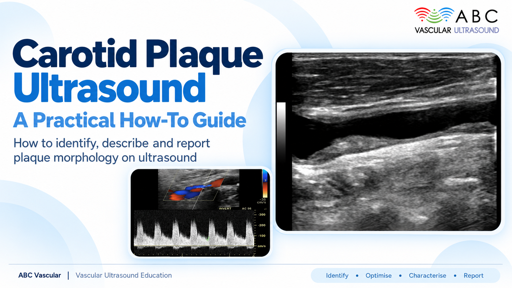
Carotid Plaque Ultrasound: A Practical How-To Guide

Why plaque characterisation matters: moving beyond percentage stenosis
Step-by-step protocol: grayscale, colour and Doppler settings for assessing plaque
Reporting plaque morphology: echogenicity, surface irregularity, ulceration and what to document

4.83 out of 5 ★★★★★
on Facebook Review and
 

Carotid ultrasound is one of the most frequently requested vascular studies in clinical practice, and for good reason. It is non-invasive, repeatable, and delivers real-time information about both the degree of stenosis and the nature of the plaque causing it. Yet a stenosis percentage on its own tells only part of the story. Two patients with an apparently similar narrowing can carry very different embolic risk depending on how their plaque looks.

This practical, step-by-step guide walks through how to characterise carotid atherosclerotic plaque on ultrasound – from the settings that make subtle findings visible, to the morphology you should document in every report.

Beyond Percentage Stenosis: Why Plaque Characterisation Matters

The degree of stenosis remains central to risk stratification, and velocity criteria continue to anchor most reporting protocols. But plaque composition increasingly guides clinical decision-making. Echolucent, heterogeneous plaques with irregular or ulcerated surfaces are more often associated with symptoms and embolic events than smooth, uniformly calcified lesions – even when the measured stenosis is comparable.

Major scientific societies, including the ESVS, recognise plaque morphology as a meaningful modifier of risk. Documenting it consistently allows you to communicate a fuller picture to referring clinicians and to track change over time in your own follow-up studies.

Before You Start: Preparation

Good characterisation begins before the probe touches the neck. Ask the patient to lie supine with the head turned slightly away from the side being examined, and keep the neck relaxed. Use a high-frequency linear transducer, typically in the 5-12 MHz range, and set your depth so the vessel occupies the upper third of the image – the plaque will be larger and easier to interrogate.

Scan in both longitudinal and transverse planes. Plaque that looks unremarkable in one plane can reveal surface irregularity or a focal ulcer in another. Start with grayscale, add colour, then finish with spectral Doppler.

Step 1: Grayscale (B-Mode) Assessment

Grayscale imaging is where plaque characterisation really happens. Optimise your gain so intraluminal blood appears black and the vessel wall is sharply defined – over-gain is the single most common error, because it makes echolucent, soft plaque appear brighter and more echogenic than it truly is.

Place the focal zone at the depth of the plaque, adjust time-gain compensation so brightness is uniform across the image, and avoid excessive compression of the neck, which can distort the plaque and the lumen. Then assess the plaque itself.

Step 2: Colour Doppler Settings

Switch to colour Doppler to map flow around the plaque, but keep the colour box as small as practical – a large box lowers frame rate and blurs the very detail you are trying to see. Lower the velocity scale (PRF) until you see no aliasing in the normal common carotid artery, and use a low wall filter so slow flow near the plaque surface is not suppressed.

Colour helps you identify the residual lumen, detect flow disturbance at the plaque shoulder, and spot areas of focal colour filling that may correspond to surface ulceration.

Step 3: Spectral Doppler

Spectral Doppler quantifies haemodynamic severity rather than plaque composition, but it completes the picture. Use angle correction of 60 degrees or less, keep the sample volume in the mid-lumen, and record peak systolic velocity, end-diastolic velocity, and the internal carotid to common carotid velocity ratio. Remember that heavily calcified plaque can underestimate stenosis on colour imaging because of acoustic shadowing, so always correlate the two.

Reporting Plaque Morphology: What to Document

A structured report makes your findings reproducible and useful. Include the following.

  • Echogenicity – describe the plaque as echolucent (soft), mixed, or echogenic (calcified), and note whether it is uniform or heterogeneous. Echolucent and heterogeneous plaques carry higher risk.
  • Surface – smooth versus irregular. Surface irregularity is a recognised marker of vulnerability.
  • Ulceration – a focal, discrete depression in the plaque surface with flow within it. Report it explicitly when present.
  • Thickness and extent – maximum plaque thickness, and whether it involves the common carotid, the bulb, or the internal carotid origin.
  • Location and laterality – always state the side and the segment.

Common Pitfalls to Avoid

  • Over-gaining the image, which falsely brightens soft plaque.
  • Correcting the Doppler angle beyond 60 degrees and overestimating velocity.
  • Calling ulceration on a noisy or single-plane image – confirm it in two planes.
  • Ignoring acoustic shadowing from calcification when estimating stenosis.
  • Reporting the stenosis percentage without any comment on plaque morphology.

From Assessment to Confident Practice

Characterising carotid plaque well is a skill built through repetition and structured learning. Once grayscale, colour, and Doppler settings become second nature, you will spot the features that change management far more reliably – and your reports will give referring clinicians the detail they need.

ABC Vascular’s accredited CME/AMA online courses take you through carotid assessment step by step, with video lectures, real-life case studies, and interactive quizzes you can complete anywhere, at your own pace. The Vascular Ultrasound Book 2025 – A Practical Guide on the Ultrasound Assessment of Carotid Atherosclerotic Plaques – complements the courses with a detailed, image-rich reference for the reporting bench. Invest in your future: master vascular ultrasound, anywhere, anytime.


Key Takeaways

  • Characterising plaque means moving beyond percentage stenosis to describe composition and surface.
  • Grayscale is the foundation; avoid over-gain and place the focus at plaque depth.
  • Keep the colour box small and the PRF low for accurate flow mapping.
  • Use spectral Doppler for haemodynamic severity, with angle correction of 60 degrees or less.
  • Document echogenicity, surface, ulceration, thickness, and location in every report.
Share this article

related courses

Practical Lower Limb Arterial Ultrasound: From Anatomy to Diagnosis

Learn how to perform a complete lower limb arterial Doppler ultrasound examination, from detailed vascular anatomy to diagnostic interpretation. This course covers the full spectrum of lower limb arterial ultrasound using high-quality videos, structured slides, and interactive quizzes; CME credit...
Free Trial

Free Trial

Explore vascular ultrasound with free sample lessons: preview carotid, venous, AAA, DVT and subclavian steal protocols. Get a real taste of our teaching before enrolling in the full courses.

Carotid Ultrasound Course and Vertebral Arteries

Learn how to conduct an ultrasound examination of the carotid and vertebral arteries. We cover the entire spectrum of the carotid ultrasound course and vertebral artery with videos, slides, and quizzes; CME credit is available.

Corso Imaging Ultrasonografico dell’Aneurisma dell’Aorta Addominale

Impara come identificare un aneurisma dell’aorta addominale (AAA), come classificarlo con l’ecografia e come misurarne il diametro massimo.

Evaluación de la Placa Aterosclerótica Carotídea

Criterios ecográficos 2D para la caracterización de la placa.

Carotid Atherosclerotic Plaque Assessment Course

Learn how to detect and characterise a carotid plaque with ultrasound. We cover the entire spectrum of the Carotid Atherosclerotic Plaque Assessment with videos, slides, and quizzes; CME credit is available.

Lower Limb Venous System Course

Master Lower Limb Venous System ultrasound with this course: gain crucial skills for DVT and venous insufficiency assessment. Earn a European certificate and 12 CME credits.

Abdominal Aortic Aneurysm Course

Learn to identify and manage abdominal aortic aneurysms with ultrasound. This course offers essential skills, e-booklets, videos, and 7 CME credits for healthcare professionals.

Screening for AAA during TTE Course

Learn how to detect an abdominal aortic aneurysm during a transthoracic echocardiography examination. We cover the entire spectrum of the Screening for AAA during TTE Course with videos, slides, and quizzes; CME credit is available.

Como Diagnosticar Trombosis Venosa Profunda Poplítea Femoral + CME Quiz

Aprender los principales criterios de diagnóstico para ultrasonido.

Leave a Reply

New Release

Your Ultimate Guide to Carotid plaque Ultrasound Assessment

By: C. Vitale & F. D'Abate

Explore the world of carotid atherosclerotic plaques with ABC Vascular’s latest eBook, “A Practical Guide on the Ultrasound Assessment of Carotid Atherosclerotic Plaques”. This guide offers healthcare professionals a comprehensive understanding of carotid plaque ultrasound assessment and its role in cardiovascular risk management.

Trusted by

FREE EBOOK · 2027 EDITION

Before you go,
take the guide.

Explore five clinical challenges in vascular ultrasound with our free illustrated eBook.

Get my free eBook

15 illustrated pages · English PDF

Vascular Ultrasound: From images to decisions, 2027 edition