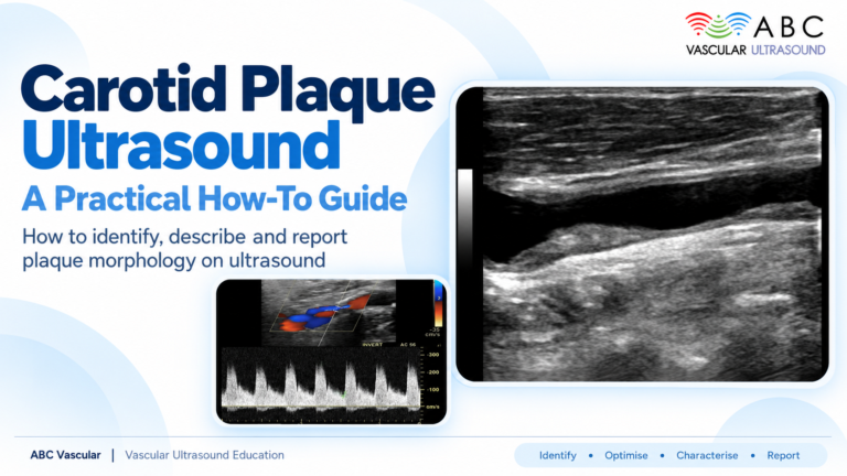

Patient-journey framework article: opens by framing five recurring clinical questions ('Is disease present?', 'How severe?', 'Haemodynamically significant?', 'Progression?', 'Has treatment worked?'), then works through carotid, peripheral arterial, venous, aortic and other territories (renal, mesenteric, upper limb) with practical examples at each journey stage. Covers advantages, limitations and operator dependence, when CTA/MRA/angiography is still needed, key takeaways, FAQs, and a restrained CTA to abcvascular.com.
 and
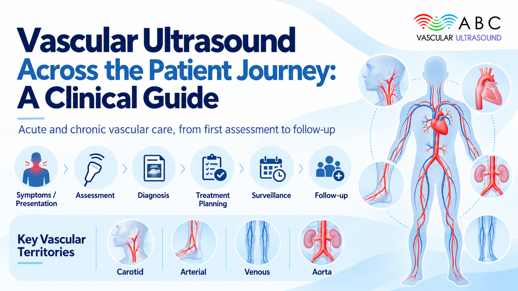
A patient rarely arrives with a diagnosis. They arrive with a symptom: a transient visual disturbance, a calf that cramps after 200 metres, a swollen leg, an incidental pulsatile abdominal mass. Deciding what happens next is where vascular imaging proves its worth. A vascular ultrasound scan can follow that patient from first presentation through diagnosis, triage, treatment planning and long-term surveillance, answering a slightly different clinical question at every stage.
Vascular ultrasound is rarely a single event. It is a thread running through an episode of care. The same non-invasive, real-time technique that confirms a deep vein thrombosis on Monday may, six months later, document that a vein has recanalised or that an aneurysm has grown by 4 mm. That continuity – the ability to repeat a safe study on the same anatomy and compare it directly – is what makes duplex ultrasound so useful to clinicians, sonographers, vascular scientists and referring physicians alike.
Across every territory, vascular imaging tends to be requested to answer one of five questions. Framing a referral around the question, rather than the vessel alone, sharpens the study and the report.
This is the screening and first-line question. A DVT ultrasound looks for non-compressibility of a deep vein; a carotid ultrasound screens for plaque in a patient after a TIA. Here the technique is chosen for availability, safety and speed, and a normal result is genuinely reassuring.
Severity means measuring, not just detecting. A stenosis is graded by peak systolic velocity and its ratio to the pre-stenotic segment; an aneurysm is measured in two planes; plaque is described by thickness, echogenicity and surface. Numbers allow comparison over time.
Anatomy alone does not settle management. Doppler ultrasound adds the haemodynamic dimension – flow direction, waveform shape, spectral broadening, post-stenotic turbulence – so a lesion can be judged by its physiological effect, not just its appearance.
Chronic vascular disease is followed, not cured. Serial surveillance detects a growing abdominal aortic aneurysm, a tightening carotid stenosis or a failing bypass before symptoms return.
Follow-up after intervention is a core role: confirming patency after carotid endarterectomy, checking a stent or vein graft, or documenting resolution after anticoagulation for venous thrombosis.
The value of vascular ultrasound becomes concrete when it is applied to specific vessels and specific decisions.
After a transient ischaemic attack, fast access to carotid ultrasound supports triage. A haemodynamically significant stenosis may prompt urgent intervention, while a plaque that is echolucent, irregular or ulcerated raises concern beyond the degree of narrowing alone. In surveillance, serial carotid ultrasound tracks progression in a patient managed medically – answering all five questions in a single, repeatable study.
For peripheral arterial disease ultrasound, the ankle-brachial index is the first filter, but duplex ultrasound maps where disease sits and whether it is significant – useful for planning angioplasty or bypass. In acute limb ischaemia, bedside assessment can distinguish an in-situ thrombosis from embolus and show whether a vessel is occluded or merely low-flow. After revascularisation, arterial ultrasound follow-up detects restenosis or a failing graft while it is still salvageable, when intervention is still straightforward.
Suspected DVT is one of the highest-volume referral questions, and DVT ultrasound – compression, with colour Doppler where needed – remains the diagnostic standard for proximal thrombus. Superficial venous thrombosis is characterised by extent and proximity to the deep system, guiding whether anticoagulation is warranted. In chronic venous disease, duplex ultrasound identifies reflux and obstruction and documents post-thrombotic changes, informing decisions on compression, ablation or stenting.
Abdominal aortic aneurysm is where serial monitoring is most clearly embedded in practice. Programme-based AAA screening measures aortic diameter; surveillance intervals then scale with size, because growth rate, not any single number, drives the decision to repair. After endovascular or open repair, aortic ultrasound assesses the sac, graft patency and endoleak alongside cross-sectional imaging, and a suprarenal AAA is measured where the visible aortic segment allows.
The same questions apply elsewhere. Renal artery duplex ultrasound screens for renovascular disease in selected patients with hypertension or deteriorating renal function. Mesenteric Doppler ultrasound can support the assessment of chronic mesenteric ischaemia, where post-prandial flow changes are informative. In the upper limb, ultrasound evaluates dialysis access, thoracic outlet compression and subclavian disease, areas where a non-invasive, repeatable study is especially welcome.
The reasons it is so widely used are practical. It is non-invasive and uses no ionising radiation, so it is safe to repeat in pregnancy, in children, in young patients and in those needing frequent follow-up. It is real-time and gives both anatomical and haemodynamic information in one study. It can be performed at the bedside in a symptomatic or unstable patient, and because it is repeatable it is well suited to serial monitoring and to comparing findings against a previous baseline.
Vascular ultrasound is not a universal answer, and claiming otherwise undermines the credibility of the referral. It is operator dependent: image quality and interpretation vary with skill, technique and equipment. Access can be limited by body habitus, bowel gas, vessel depth, overlying calcification or recent surgery. It is a focused, segmental technique, so a full anatomical map may not emerge from one study. When calcium obscures a vessel, when a complete mapping of the aortoiliac tree is required before complex intervention, or when the aorta, mesenteric vessels or distal runoff must be assessed in full, CTA, MRA or catheter angiography may still be the better choice. Ultrasound and cross-sectional vascular imaging are complements, not rivals.
Vascular ultrasound is a journey-long tool, not a one-off test, supporting diagnosis, triage, planning, surveillance and follow-up.
Framing a referral around a specific clinical question improves both the study and the report.
Duplex and Doppler ultrasound combine anatomical detail with haemodynamic information, which is central to judging significance.
It is non-invasive, radiation-free, real-time and repeatable, making it ideal for serial monitoring.
Limitations – operator dependence, limited acoustic access and segmental views – should be stated honestly, and CTA, MRA or angiography used when they add value.
No. It is non-invasive, uses no ionising radiation and involves no needles or contrast, so it can be repeated safely when serial monitoring is needed.
Often it can answer the clinical question on its own, but it does not replace angiography in every setting. Complex pre-procedural mapping or vessels obscured by calcium are common triggers for CTA, MRA or catheter angiography.
The interval depends on the disease and its severity – an aneurysm is rescanned according to size and growth rate, whereas a treated stenosis may be followed on a fixed schedule. Intervals follow local protocols.
It adds blood flow. Doppler interrogation shows direction, velocity and waveform, allowing a lesion to be judged by its haemodynamic effect rather than its appearance alone.
Vascular ultrasound rewards technique and interpretation as much as equipment. The clinicians and sonographers who get the most from each scan are those who know exactly which question they are answering and which haemodynamic markers will answer it. If you would like to build that confidence, explore the accredited vascular ultrasound courses at abcvascular.com, where practical, peer-level training covers each territory from probe position to reporting.