

Case Studies
Access a wide variety of real life case studies ranging from basic to more complex vascular pathologies. Solve challenging cases and get virtual experience of vascular ultrasound.
Interpreting Doppler Waveforms
Doppler waveform analysis is crucial for diagnosing arterial and venous diseases. This summary highlights the SVM and SVU's consensus on standardizing nomenclature for better patient care.
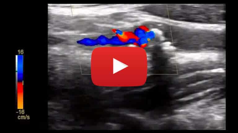
Occlusion of the Common Carotid Artery: Collateral Pathway
Case study description: Occlusion of the CCA Video length: 2 mins Audio: Yes, with voice-over explanation Jump to video below > Retrograde filling of the external carotid artery (ECA) supplying antegrade flow into the internal carotid artery (ICA) in the presence of…

Assessment of Carotid Arterial Plaque
Discover the latest ASE recommendations on using ultrasound to assess carotid plaque for atherosclerosis characterization and cardiovascular risk evaluation.

Free Floating Distal Thrombus Tongue
This short video demonstrates a case of acute deep vein thrombosis (DVT) of the femoral vein with evidence of a mobile tongue of thrombus extending and floating within the distal common femoral vein.
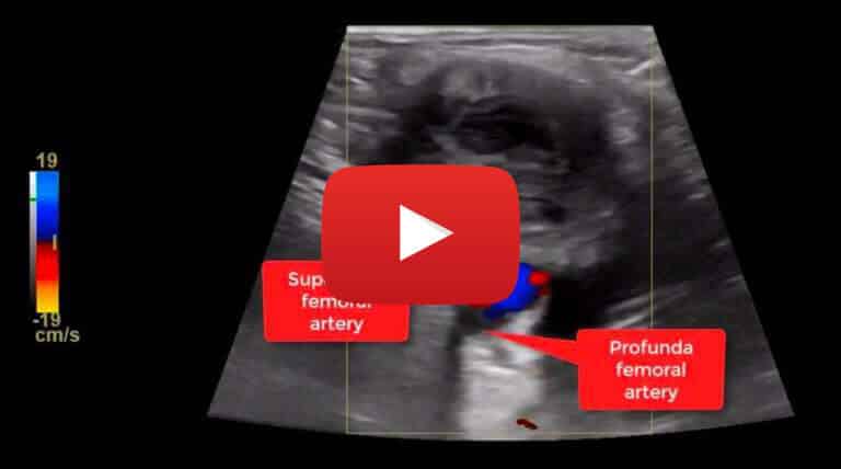
A Thrombosed Common Femoral Artery Pseudoaneurysm: Ultrasound Features
Case study description: Short video showing the features of a thrombosed common femoral artery pseudoaneurysm Video length: 40 secs Audio: No audio Jump to video below > A large perfused pseudoaneurysm of the common femoral artery (CFA) was detected with…
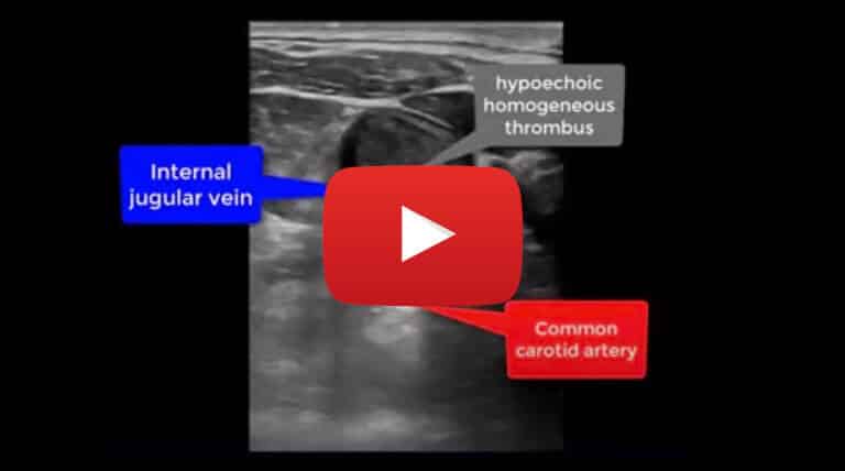
Acute Deep Vein Thrombosis of the Internal Jugular Vein: B-Mode Imaging
Case study description: Short video showing acute DVT of the IJV Video length: 33 secs Audio: No audio This video shows a hypoechoic thrombus attached to the anterior wall of the internal jugular vein with the remaining part of the…
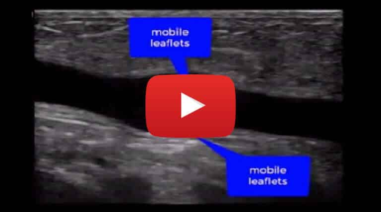
Normal vs Abnormal Venous Valve
Case study description: Short video showing a normal vs an abnormal venous valve Video length: 35 secs Audio: No audio Jump to video below > Venous valves are bicuspid valves that allow venous blood to flow in one direction only…
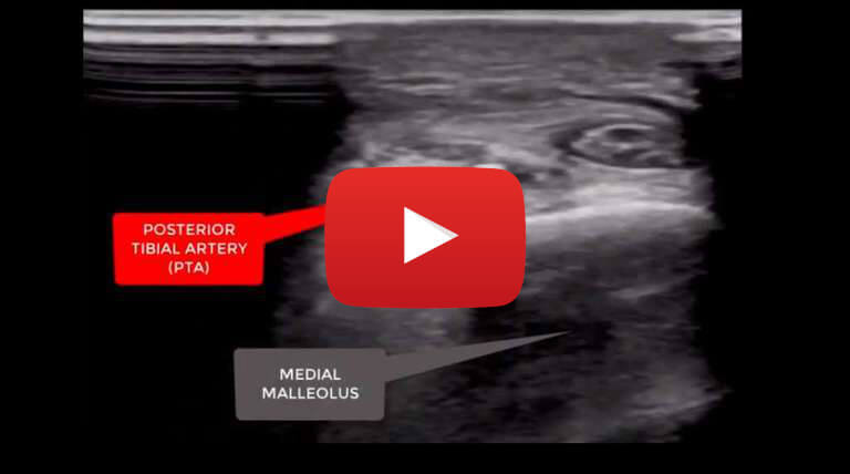
Calcified Occlusion of the Posterior Tibial Artery: Tips for Ultrasound Operators
Case study description: Short video with tips on viewing calcified occlusion of the posterior tibial artery Video length: 2 mins Audio: Yes, with voice-over explanation Jump to video below > This is a case of a 58 years old male…
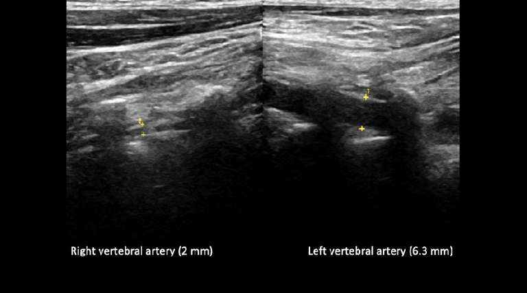
Vertebral Artery Hypoplasia: Ultrasound Appearance and Criteria
Explore the ultrasound criteria and appearance of Vertebral Artery Hypoplasia (VAH), a condition often seen in patients with posterior circulation stroke, and understand its clinical implications.
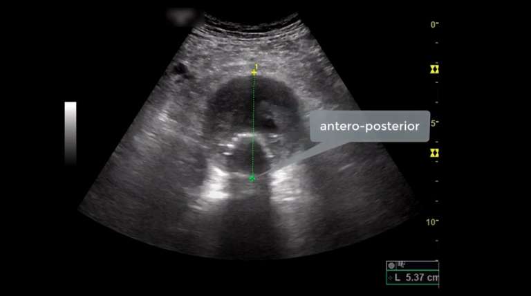
Normal Colour Doppler Flow Appearance of an AAA After Endovascular Repair (EVAR)
Ultrasound is key for assessing abdominal aorta post-EVAR, focusing on residual sac size, detecting endoleaks, and ensuring stent-graft patency. Learn the importance of color Doppler in EVAR evaluation.


