Calcified Occlusion of the Posterior Tibial Artery: Tips for Ultrasound Operators
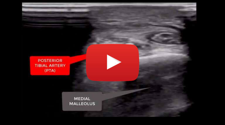
Case study description: Short video with tips on viewing calcified occlusion of the posterior tibial artery
Video length: 2 mins
Audio: Yes, with voice-over explanation
This is a case of a 58 years old male patient with a 30 years history of type 1 diabetes in treatment with insulin and metformin.
Patient was referred to the vascular laboratory for a lower limb arterial ultrasound examination due to the presence of necrotic second toe of the right lower limb.
The lower limb arterial ultrasound assessment demonstrated on the right side, patency of the femoral and popliteal segments in presence of diffuse non-haemodynamic hyperechoic plaques and no evidence of focal stenoses.
The tibio-peroneal trunk, peroneal artery and anterior tibial artery resulted patent with diffuse hyperechoic plaques and good biphasic flow. The posterior tibial artery appeared of small calibre, chronically occluded with occlusive hyperechoic plaques. Patients with diabetes are often found to have diffuse calcifications throughout the below knee arteries.
Determining the patency of the tibial arteries and the more distal segments (such as the common plantar artery and the dorsalis paedis artery) is of paramount importance in this cohort of patients considering the high risk to develop wound healing complications and consequent amputations.
In order to improve the visualisation of the distal tibial arteries there are two main tips that the ultrasound operators should be aware of and these are:
- In the more superficial course of the tibial arteries the use of a high frequency array (18-5MHz) can be of help as it improves the lateral resolution and allows for a better visualisation of the intraluminal atherosclerotic disease as shown in this video.
- Colour Doppler flow should be optimised in order to detect low velocity flow. A low colour flow scale is therefore needed alongside an increase of the overall colour Doppler flow gain.
Power angio, contrast enhanced ultrasound and B-flow imaging may reveal to be useful to detect low velocity flow, however not all ultrasound operators may have access to such technologies.


